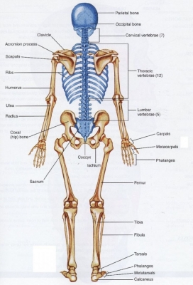
From the back you can clearly see the complex structure of the spinal column and paired ribs. Each wing-like shoulder blade forms part of the shoulder gridle, where the arm attaches to the axial skeleton.
Suture
Suture lines form where bones of the skull have joined (fused) together. A tiny amount of movement is permitted at sutures, which contributes to the compliance and elasticity of the skull. These joints are synarthroses. It is normal for many of the bones of the skull to remain unfused at birth.
Parietal bones
A pair of bones makes up the sides of the skull. The parietal bones are marked internally by meningeal blood vessels and externally by the temporal muscles. They meet at the top of the head (sagittal suture) and form a roof for the cranium.
Occipital bone
This flattish bone forms the back of the skull. The occipital is cupped like a saucer in order to house the back part of the brain. It is one of seven bones that fuse together to form the skull and is directly next to five of the cranium bones.
Atlas
The uppermost vertebra of the spine, this bone supports the head. The atlas does not look like a typical vertebra, with its ring-like structure and the absence of a body, which is actually fused to the axis. Other anatomical landmarks on the atlas include the anterior arch and tubercle, posterior arch and tubercle, vertebral notches, facets, and transverse processes.
Axis
The second bone of the vertebral column allows the head to move from side to side. The axis is somewhat analogous to the other cervical vertebrae in shape, but it differs slightly for two reasons: its spinous process isn’t as obviously bifid, and the presence of the dens. The spinous process serves as the attachment site for many muscles of the spine, particularly those close to the skull, as well as the nuchal ligament.
Cervical vertebrae
These seven bones form the upper part of the spine. Among the vertebrae of the spinal column, the cervical vertebrae are the thinnest and most delicate bones. Yet, in spite of their size, the cervical vertebrae have the huge jobs of supporting the head, protecting the spinal cord, and providing mobility to the head and neck.
Acromion
This is the highest part of the shoulder blade. It is an important landmark of the skeletal system and a muscle attachment point essential to the function of the shoulder joint. The acromion also forms the acromioclavicular (AC) joint with the clavicle.
Clavicle
This long bone is called the collarbone. The clavicle (collarbone) extends between the manubrium of the sternum and the acromion of the scapula. It is classed as a long bone, and can be palpated along its length. In thin individuals, it is visible under the skin.
Scapula
Also called the shoulder blade, it connects the arm to the shoulder. In humans they are triangular and lie on the upper back between the levels of the second and eighth ribs. A scapula’s posterior surface is crossed obliquely by a prominent ridge, the spine, which divides the bone into two concave areas, the supraspinous and infraspinous fossae.
Spinal column
Also called the backbone, it is made up of 24 small bones (vertebrae), which protect the nerves. The Spinal Column is also called the vertebral column. The bones in the spine are called vertebrae (ver-ta-bray). The column starts at the base of the skull and continues to the pelvis. Alternate layers of bone (vertebrae) and cartilage (car-til-ledge, the intervertebral discs) stack vertically one on top of the other in the spinal column. The lattice-like structure of the cancellous bone (cancel-lus, the spongy interior) in a vertebra absorbs external pressure.
Rib
The 12 pairs of curved rib bones protect the heart and lungs. The ribs partially enclose and protect the chest cavity, where many vital organs (including the heart and the lungs) are located. The rib cage is collectively made up of long, curved individual bones with joint-connections to the spinal vertebrae.
Humerus
This is the upper arm bone. It is located between the elbow joint and the shoulder. At the elbow, it connects primarily to the ulna, as the forearm’s radial bone connects to the wrist.
Lumber vertebrae
These five bones form the lower part of the spinal column. These vertebrae carry all of the upper body’s weight while providing flexibility and movement to the trunk region. They also protect the delicate spinal cord and nerves within their vertebral canal.
Ulna
This is the inner bone of the forearm. The ulna is located on the opposite side of the forearm from the thumb. It joins with the humerus on its larger end to make the elbow joint, and joins with the carpal bones of the hand at its smaller end.
Ilium
The ilium is one of the bones that makes up the pelvis. In humans, it is divided into two sections: the body and the ala, indicated by a line on the surface of the bone. The other two bones that form the fused pelvis are the ischium and the pubis, which lie below the ilium.
Radius
This is the outer bone of the forearm. It lies laterally and parallel to ulna, the second of the forearm bones. The radius pivots around the ulna to produce movement at the proximal and distal radio-ulnar joints.
Sacrum
This is large, triangular bone that forms the base of the spine. It forms the solid base of the spinal column where it intersects with the hip bones to form the pelvis. The sacrum is a very strong bone that supports the weight of the upper body as it is spread across the pelvis and into the legs.
Coccyx
Several tiny bones at the end of the spine fuse to form the coccyx. It is composed of three to five coccygeal vertebrae or spinal bones. The vertebrae may be fused together to form a single bone; however, in some cases, the first vertebra is separate from the others.
Femur
The femur, or thigh bone, is the longest bone in the body. It functions in supporting the weight of the body and allowing motion of the leg. The femur articulates proximally with the acetabulum of the pelvis forming the hip joint, and distally with the tibia and patella to form the knee joint.
Femoral condyles
These rounded, knobbly ends of the femur form part of the knee joint. There are two condyles on each leg known as the medial and lateral femoral condyles. If there is a fracture (break) in part of the condyle, this is known as a fracture of the femoral condyle. Physiotherapy is very important during the rehabilitation following a femoral condyle fracture.
Tibia
The front of this bone is the shin. It forms the knee joint with the femur and the ankle joint with the fibula and tarsus. Many powerful muscles that move the foot and lower leg are anchored to the tibia.
Fibula
The smaller bone of the lower leg, this is located alongside the tibia. It runs parallel to the tibia, or shin bone, and plays a significant role in stabilizing the ankle and supporting the muscles of the lower leg. Compared to the tibia, the fibula is about the same length, but is considerably thinner.
Heel bone
The largest bone in the foot, this is also called calcaneus. It is situated in the back of the foot, just below the talus, tibia, and fibula bones of the lower leg. Of all of the bones in the foot, the heel bone is the largest.
Picture Credit : Google