The respiratory system is a vast network of millions of airways, spreading like the branches of a tree into the lungs. With each breath, air sucked in through the nose or mouth rushes down the windpipe, or trachea. This carries air to a fork deep inside the chest where the airways divide in two. One of the branches, or bronchi, leads to the left lung, while the other leads to the right. Air comes in when the lungs expand, and is pushed out when they shrink back.
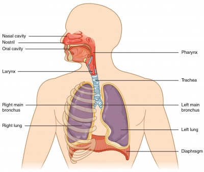
Nasal cavity
The nasal cavity anatomy is essential for both breathing and our sense of smell (olfaction). But did you know that 80% of taste actually comes from what we smell? That is why food is almost tasteless when our nose is clogged. Air that enters through the nose is warmed and filtered in this space.
Nostril
A nostril is one of the two channels of the nose, from the point where they bifurcate to the external opening. Air enters the body through the twin openings in the nose.
Mouth
The mouth, also known as the oral cavity, is the secondary external opening for the respiratory tract. Most normal breathing takes place through the nasal cavity, but the oral cavity can be used to supplement or replace the nasal cavity’s functions when needed. Air is also breathed in through the mouth.
Pharynx
Pharynx, cone-shaped passageway leading from the oral and nasal cavities in the head to the esophagus and larynx. The nose and mouth are linked to the larynx by this airway.
Epiglottis
The epiglottis is a cartilaginous flap that extends in front and above the laryngeal inlet, or more specifically the rima glottidis (glottis). Food or liquid is prevented from entering the trachea by this flap of cartilage.
Larynx
This is the top part of the trachea. It has bands, called vocal cords, which contract and relax to create sounds.
Trachea
The windpipe, a strong tube of muscle and rings of cartilage, carries air from the larynx to the lungs.
Intercostal muscles
The intercostal muscles are a group of muscles found between the ribs which are responsible for helping form and maintain the cavity produced by the ribs. During breathing muscles between the ribs pull the rib cage up and down.
Ribcage
The human rib cage is a component of the human respiratory system. It encloses the thoracic cavity, which contains the lungs. An inhalation is accomplished when the muscular diaphragm, at the floor of the thoracic cavity, contracts and flattens, while the contraction of intercostal muscles lift the rib cage up and out. These bones surround and protect the lungs.
Left lung
The left lung has just two lobes. The lobes are made of sponge-like tissue that is surrounded by a membrane called pleura, which separates the lungs from the chest wall. This is the smaller lung because it shares space with the heart.
Bronchi
Bronchi are plural for bronchus and represent the passageways leading into the lungs. These two branches (one is a bronchus) lead from the trachea into each lung.
Bronchioles
The bronchioles are tubes in the lungs which branch off from the larger bronchi that enter each lung, from the large and singular trachea which connects to the mouth. The tiniest branches at the end of the air passages are finer than hairs.
Right lung
This is the larger of the two lungs. It has three sections called lobes.
Heart
The heart pumps blood to the lungs to pick up oxygen. The heart and lungs work together to make sure the body has the oxygen-rich blood it needs to function properly. The Pulmonary Loop The right side of the heart picks up the oxygen-poor blood from the body and moves it to the lungs for cleaning and re-oxygenating.
Visceral pleura
This membrane covers the surface of the lungs. It is continuous with the parietal pleura at the hilum of each lung (this is where structures enter and leave the lung).
Pleural cavity
The pleural cavity is a potential space between the parietal and visceral pleura. It contains a small volume of serous fluid, which has two major functions. A thin layer of fluid lies between the visceral and parietal plurae to lubricate lung movement.
Parietal pleura
The parietal pleura is sensitive to pressure, pain, and temperature. This membrane lines the inner wall of the chest.
Diaphragm
The diaphragm, located below the lungs, is the major muscle of respiration. It is a large, dome-shaped muscle that contracts rhythmically and continually, and most of the time, involuntarily. To increase space in the chest and draw air into the lungs, this sheet of muscle contracts and flattens.
Picture Credit : Google