The average eyeball is about 2.5 cm (1 in) in diameter and is made up of two fluid-filled cavities – a small space in front of the lens and a larger area behind it. Light enters the eye through the pupil, which is an adjustable window between the cornea and the lens.
Superior rectus muscle
This muscle controls upward movements. This group of muscles serves to move the eyes within the orbit. It includes the superior rectus, inferior rectus, medial rectus, lateral rectus, superior oblique and inferior oblique muscles.
Lateral rectus muscle
Lateral rectus muscle is one of the 4 straight muscles of the orbit responsible for the movement of the eye in the cardinal directions. The eye is pulled from side to side by this muscle.
Superior oblique muscle
Superior oblique is the longest muscle in this group, spanning from the body of sphenoid bone to the super lateral aspect of the eyeball. This rotates the eye upwards and towards the nose.
Inferior oblique muscle
Like the other eye muscles, inferior oblique is named by its position within the orbit, relative to the eyeball. This muscle rotates the eye downwards and towards the outside of the head.
Inferior rectus muscle
The inferior rectus muscle originates from the common tendinous ring, and goes on to attach at the lower anterior surface of the eyeball. The eye is pulled downwards by this muscle.
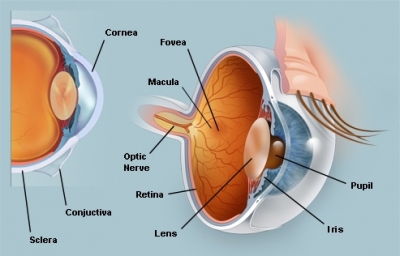
Sclera
The outer coating is also called the white of the eye. In fact, the sclera forms more than 80 percent of the surface area of the eyeball, extending from the cornea all the way to the optic nerve, which exits the back of the eye. Only a small portion of the anterior sclera is visible.
Retina
This layer contains millions of cells that detect light. The purpose of the retina is to receive light that the lens has focused, convert the light into neural signals, and send these signals on to the brain for visual recognition.
Fovea
The central part of the retina, this contains colour-detecting cones. The fovea is responsible for sharp central vision (also called foveal vision), which is necessary in humans for activities for which visual detail is of primary importance, such as reading and driving. The fovea is surrounded by the parafovea belt and the perifovea outer region.
Vitreous humour
The vitreous humour (also known simply as the vitreous) is a clear, colourless fluid that fills the space between the lens and the retina of your eye. 99% of it consists of water and the rest is a mixture of collagen, proteins, salts and sugars. This is a thick jelly that fills the back of the eye.
Pupil
The pupil is the opening that allows light into the eye. While your two pupils will usually be roughly the same size, pupil size overall can fluctuate. Factors that cause your pupils to become bigger or smaller are light (or the lack of it), certain medications and disease, and even how mentally interesting or taxing you find something.
Cornea
A clear layer, the cornea helps to focus light. Your cornea can also filter out some of the sun’s ultraviolet light. But not much, so your best bet to keep it health is to wear a pair of wraparound sunglasses when you’re outdoors.
Lens
This structure changes shape to focus light on the retina. In other words, it focuses the light rays that pass through it (and onto the retina) in order to create clear images of objects that are positioned at various distances. It also works together with the cornea to refract, or bend, light.
Iris
The iris is a circle of muscle that controls how much light enters the eye. Together with the pupil, the iris is responsible for regulating the amount of light that gets into the eye. Too much or too little light can hamper vision.
Ciliary muscles
The ciliary muscle occupies the biggest portion of the ciliary body, which lies between the anterior border of the choroid and iris. These contract or relax to adjust the shape of the lens.
Optic nerve
The optic nerve is located in the back of the eye. It is also called the second cranial nerve or cranial nerve II. It is the second of several pairs of cranial nerves. Signals from receptors in the retina are carried to the brain along this nerve.
Light detectors
Rods and cones are two types of light receptor cell on the retina. Rods pick up dim light, while cones detect colour and detail. Then they send information about what they record to the brain via the optic nerve.
Picture Credit : Google