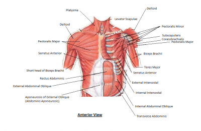
The muscles of the chest help with the process of breathing. They pull the ribs up and out, making more space for the lungs to expand as we breathe in. When they relax, the space gets smaller and the air is forced out again.
Clavicle
The clavicle is an elongated, S-shaped bone that rests horizontally at the sternum across the upper part of the ribcage, and the acromial end of the scapula. This is also called collarbone.
Pectoralis major
The largest chest muscle, it is attached to the sternum, clavicle, humerus (upper arm bone), and ribs. The pectoralis major has a broad origin, based on which it is divided into three parts: clavicular part, sternocostal part and abdominal part. All three parts converge laterally and insert onto the greater tubercle of humerus.
Sternum
The sternum is the bone that lies in the anterior midline of our thorax. It forms part of the rib cage and the anterior-most part of the thorax. Several muscles attach to the sternum, or breastbone.
Deltoid
The deltoid is a thick, triangular shoulder muscle. It gets its name because of its similar shape to the Greek letter ‘delta’ (?). Covering the shoulder joint, this muscle raises the arm.
Serratus anterior
The serratus anterior muscle is a fan-shaped muscle at the lateral wall of the thorax. Its main part lies deep under the scapula and the pectoral muscles. This muscle connects to the upper ribs.
Costal cartilage
Tough, springy tissue connects the sternum to the ribs. The costal cartilage is segments of cartilage that connect the sternum to the ribs and help to extend the ribs into a forward motion. This cartilage also contributes to elasticity within the walls of the thorax, allowing the chest to expand during respiration
Rectus abdominis
Rectus abdominis, informally known as the abs muscle, is a long muscle of the anterior abdominal wall. Attached to the lower sternum and coastal cartilages, this helps keep the body upright.
External oblique
External abdominal oblique is a paired muscle located on the lateral sides of the abdominal wall. Along with internal abdominal oblique and transversus abdominis, it comprises the lateral abdominal muscles. This outer muscle layer helps to force air out of the lungs when we breathe out.
Rib
Each rib is thin and curved, with an inside groove for veins, arteries, and nerves. In most tetrapods, ribs surround the chest, enabling the lungs to expand and thus facilitate breathing by expanding the chest cavity.
Internal oblique muscles
Internal abdominal oblique is a broad thin muscular sheet found on the lateral side of the abdomen. These help to push air out of the lungs when we breathe out.
Intercostals muscles
Between the ribs are three layers of intercostals muscles (“intercostals” simply means “between the ribs”). The muscle fibres run in different directions so the ribs can be pulled in different ways.
Internal muscle
Internal intercostals are part of the muscles of the thoracic wall located in the intercostal spaces between the ribs. When you breathe out, this muscle pulls the ribcage down and out.
External muscle
The external intercostal muscles are the most superficial set of muscles that occupy the 11 intercostal spaces. This pulls the ribcage up and out when breathing in.
Innermost muscle
Innermost intercostals comprise the third and deepest layer of intercostal muscles. This muscle lowers the ribs when breathing out.
Picture Credit : Google
