A healthy adult’s heart is about the size of a clenched fist. It sits in the thorax (chest) and in most people the tip points towards the left side of the body.
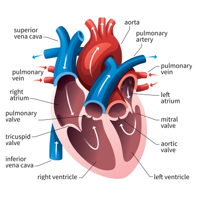
Aorta
The aorta is the first segment of the systemic arterial circulation, originating directly from the left ventricle of the heart. The body’s main artery carries blood from the heart to the rest of the body.
Superior vena cava
The superior vena cava (SVC, also known as the cava or cva) is a short, but large diameter vein located in the anterior right superior mediastinum. This large vein returns oxygen-poor blood from the upper body to the heart.
Pulmonary artery
The pulmonary arteries and the pulmonary veins are the vessels of the pulmonary circulation; which means they are responsible for carrying the oxygenated blood to the heart from the lungs and carrying the deoxygenated blood from the heart to the lungs. Blood is carried to the lungs by this vessel.
Coronary artery
The heart’s own blood supply is delivered by this artery. The right coronary artery supplies blood mainly to the right side of the heart. The right side of the heart is smaller because it pumps blood only to the lungs.
The left coronary artery, which branches into the left anterior descending artery and the circumflex artery, supplies blood to the left side of the heart.
Muscle structure
Heart muscle shows that the structure is a network of interlocking fibres. The oval discs are mitochondria, which supply the muscle cells with the energy they need.
Pericardium
This tough, double-layered “bag” around the heart keeps infections out and stops the heart from expanding too much when blood flows into it. It also allows the heart to beat without rubbing against other organs.
Picture Credit : Google