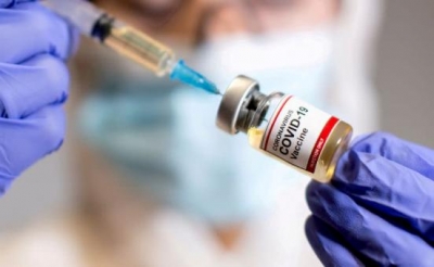
Why are the Covid-19 vaccines a major milestone?

The Covid-19 disease is caused by a strain of positive-sense single-stranded RNA virus called Severe Acute Respiratory Syndrome Coronavirus 2, also known as SARS-CoV-2. It is responsible for the ongoing global coronavirus pandemic which has killed over 38 lakh people since it was first detected in December, 2019.

Vaccines are critical in the fight against Covid-19, along with safety measures such as proper hand and face hygiene, the use of face masks and social distancing.

Some of the vaccines approved by the World Health Organization as of June 2021 are as follows: AstraZeneca/Oxford vaccine, Johnson and Johnson, Moderna, Pfizer/BionTech, Sinopharm, Sinovac.

Covaxin, India’s first indigenous Covid-19 vaccine has been developed by Bharat Biotech in collaboration with the Indian Council of Medical Research and the National Institute of Virology.

The AstraZeneca Vaccine is being produced in India as Covishield. The Russian vaccine Sputnik V has also been approved for use in India amid the second wave of the pandemic.

The Covid-19 vaccines are safe for people above 18 years and older, including senior citizens and people with pre-existing conditions like hypertension, diabetes and asthma. As of now, Covid-19 vaccine trials for children have begun in the USA, Singapore, Japan, and parts of Europe. China has approved Sinovac and Sinopharm vaccines for children as young as 3-years and above. In India too, clinical trials for children have begun at AIIMS, Delhi.

Picture Credit : Google

Why is robotic surgery so precise?

In robotic surgery, a robotic system responds to the movements of the surgeon’s hands to perform complex surgeries, using miniaturized instruments and a three-dimensional camera.

It allows the doctor to perform minimally invasive procedures involving tiny incisions with more precision than possible conventionally. Typically a robotic surgical system will include a camera arm and mechanical arms with attached surgical instruments.

To understand better, think of video games. One can control the movements in the game using the console or keys available. In robotic surgery too, the surgeon performs precise, delicate movements using master controls while sitting at a nearby console.

The advantage of this type of surgery is that the surgeon is able to see a high-definition, magnified, 3-D view of the surgical field and has greater control and dexterity over the procedure. Also, the patient has a lesser risk of infection, smaller scars, lesser blood loss and a shorter hospital stay.

Picture Credit : Google

What is the Human Genome Project?

The Human Genome Project (HGP) was an international, collaborative research effort to determine the DNA sequence of the entire human genome (the complete genetic material present in the human organism). The project was launched in 1990 and was declared complete in 2003.

The HGP researchers deciphered the human genome in three major ways: determining the order or sequence of all the bases in our genome’s DNA; making maps that show the locations of genes for major sections of all our chromosomes; and producing what are called linkage maps, through which inherited traits (such as those for genetic disease) can be tracked over generations.

The HGP revealed that the human genome contained more than 2.85 billion nucleotides and that there are approximately 22,300 protein - coding genes in human beings.

The HGP has given the world a resource of detailed information about the structure, organization and function of the complete set of human genes.

As researchers learn more and more about the functions of genes and proteins, this knowledge is all set to have an even greater impact in the fields of medicine, biotechnology and life sciences.

Picture Credit : Google

What is cloning and what are its implications?

Cloning is the process of generating a genetically identical copy of a cell or organism. The closest that we can see this occurring naturally, is in identical twins.

Scientists use two methods to get an exact, genetic copy of an organism. In the first, known as Artificial Embryo Twinning, scientists mimic the natural process of creating identical twins. The only difference being that in the natural process twinning takes place inside the womb and in the artificial twinning method, the splitting of the fertilized egg into two genetically identical embryos takes place in the lab.

The second method is called ‘Somatic Cell Nuclear Transfer’ or SCNT. In this process a viable embryo is created using a body cell and an egg cell.

Scientists at the Roslin Institute in Edinburgh, UK made history on 5th July 1996, when they successfully cloned a sheep named Dolly using the SCNT method.

In this process of cloning, the scientists extract the DNA from an animal cell and implant it into an egg cell taken from another animal. Before implanting it, the DNA of the recipient cell is removed.

The idea of cloning can be exciting and frightening at the same time. Once the techniques are perfected, cloning may be used in preserving endangered species and for reviving extinct species as well.

Picture Credit : Google

What is DNA profiling?

DNA profiling, also called DNA fingerprinting, is a test to identify a person’s DNA characteristics. The DNA test is called a ‘fingerprint’, because just as it is very unlikely that any two people would have the exact, same physical fingerprint, it is also very unlikely that any two people would have exactly the same DNA information.

Only a small sample of cells is needed for DNA fingerprinting. A drop of blood or strands of hair contain enough genetic material for DNA testing. The test is used to determine whether a family relationship exists between two people. It is also used to identify organisms causing a disease. It is also a forensic technique in criminal investigations and is an important and decisive component of solving crimes. For example, DNA isolated from dried blood, saliva or hair found at a crime scene can be compared to a DNA sample collected from someone suspected of a crime, to prove his or her innocence or guilt.

Picture Credit : Google

What is Jarvik 7?

You may have heard of heart transplants, where organs of diseased people are transplanted into living recipients. In many such cases there is a long list of recipients and a very short list of donors. But did you know that instead of waiting for a heart donor, there is another option for heart patients? The alternative is called the ‘Total Artificial Heart’ or TAH. The TAH is a form of mechanical circulatory support in which the patient is supported by a pneumatically powered, artificial heart.

The first TAH to be implanted was the Jarvik 7 in 1982. The Jarvik 7 was an artificial heart made of plastic and metal, named after its designer Robert Jarvik. It was the first TAH which was designed to be implanted as a permanent replacement after the natural heart was removed.

In probably one of the most dramatic surgical procedures that was ever imagined, surgeons at the University of Utah implanted the Jarvik 7 heart into Barney Clark, a 62-year-old dentist, in 1982.

Clark, who was in the final stages of heart failure, agreed to the procedure as an experiment to help medicine and did not expect to survive more than a few days. He however, went on to live for 112 days after the implantation.

With the relative success of the first operation, by the late 1980s, surgeons at 16 centers were using the Jarvik 7 as a bridge to heart transplantation. Today, though the original Jarvik 7 heart is no longer in use, this invention was truly a remarkable feat in the history of medical science.

Picture Credit : Google

What is MRI and how is it useful?

Magnetic Resonance Imaging, or MRI, is a valuable clinical and research tool. It gives a visual representation of organs and structures in the body.

Magnetic resonance imaging was made possible when Nikola Tesla first discovered the rotating magnetic field in 1882. Nearly one hundred years later in 1977, an American physician, Raymond Vahan Damadian invented the first MRI scanning machine. He was the first to perform a full body scan on a human being with a resultant diagnosis of cancer.

An MRI scanner consists of a long tube that the patient is inserted into. Here the patient is subjected to a very strong magnetic field and radio waves are applied in different directions. This causes atoms in the patient’s body to emit special signals which are detected by the MRI scanner and sent to a computer. Based on the data the computer creates an image of the part of the body to be scanned.

Although it is more expensive than other procedures, a major advantage of MRI is that dangerous X-ray radiation can be avoided. MRI’s are used to study the brain, spinal cord, bones, joints and internal organs. It is an extremely accurate diagnostic method and an invaluable tool for disease detection.

The development of MRI revolutionized the medical world. Since its discovery, MRI scans not only assist in medical procedures but also aid in research.

Picture Credit : Google

What is a CAT Scan?

A CAT scan or CT scan is a special X-ray test that produces cross-sectional or three-dimensional images of different sections of the body. The acronym ‘CAT’ stands for ‘Computerized Axial Tomography’.

The invention of computers sparked researchers to try to combine X-rays with computer technology. In 1972, a British biomedical engineer named Sir Godfrey Hounsfield, developed the CAT scan. He received the Nobel Prize in 1979 for his work.

The first clinical CT scanners began to be installed in 1974. The original systems were dedicated to taking images of the head only, but ‘whole body’ systems became available in 1976. CT scans became widely available by about 1980.

The first CT scanner took several hours for a single scan and took days to reconstruct a single image from raw data. Today, an entire chest CT of 40 slices and 8mm thickness, can be scanned in 5 to 10 seconds using the most advanced multi-slice CT system.

During its 25-year history, CT has made great improvements in speed, patient comfort and resolution. CT scanners are now used to take pictures of any part of the body and are invaluable to medical diagnosis.

Picture Credit : Google

How has heart transplant surgery changed medical history?

In a heart transplant procedure, a surgeon removes the diseased heart of a person and replaces it with a donor heart. It is used to treat the most serious cases and as an option for people in the final stages of heart failure.

This sort of surgery was unimaginable till the beginning of the 20th century. It was only the invention of the heart-lung bypass machine by John Gibbon, in 1953 that made such a procedure possible. In 1967, for the first time, a human heart from one person was transplanted into another by a surgeon named Christiaan Barnard in Cape Town, South Africa.

Dr Barnard’s team removed the heart of a 25-year-old woman, who had died following an auto accident, and placed it in the chest of Louis Washkansky, a 55-year-old man dying of heart damage. The surgery was the first of its kind and the patient survived for 18 days on the borrowed heart.

In the 1970s, with the development of better anti-rejection drugs, heart transplantation became more viable and by the late 1970s patients were living up to five years with their new hearts. Today, new approaches to heart transplant surgery are being researched, but Barnard’s operation will always be remembered as a historic achievement in the field of medicine.

Picture Credit : Google

What is polio and who was behind the polio vaccine?

Polio is caused by one of three types of poliovirus from the Enterovirus genus. It spreads through contact between people, through nasal and oral secretions and through contaminated faeces. The polio virus can damage the brain and spinal cord. In around 98 per cent of cases it is a mild attack with almost no symptoms. However in a very small percentage of people, it can cause paralysis and loss of the use of a limb.

Polio has been around for thousands of years and affected human populations at relatively low levels till the early 1900s. This was a time when other diseases like diphtheria, typhoid and tuberculosis were being treated successfully. Ironically, advances in hygiene led to the increase of polio cases. This was because in the past, infants were exposed to the polio virus through contaminated water supplies at early ages. These exposed infants developed immunity due to natural maternal antibodies still circulating in their system. However, better sanitation meant that exposure to the polio virus was getting delayed till children had lost their maternal antibodies, which led to them being more vulnerable.

By 1940, polio was a highly infectious disease and many scientists were racing to make a vaccine for it.

It was Dr Jonas Salk who finally succeeded in 1955. He started working on a vaccine against the three strains of polio and tested the vaccine on his own children first. Salk’s vaccine was composed of an injected, inactivated form of the polio virus, which retained the ability to give Immunity against the disease without the risk of infecting the patient.

However, Salk’s vaccine was not completely effective against one of the three strains of polio and it only gave immunity for a limited time. In 1957, Albert Sabin, a Polish- American medical researcher, succeeded in making an oral vaccine which used a small amount of the live virus. This vaccine became available globally in 1962 and today, the oral polio vaccine is one of the most successful vaccines of all times.

Currently, the polio virus is only found in Afghanistan and Pakistan with an occasional spread to neighbouring countries.

Picture Credit : Google

What is an endoscope and why is it important?

An endoscope is an instrument that allows a doctor to look inside the human body. Evidence of such instruments have been found since early Greek and Roman times. In 1805, Philip Bozzini made the first endoscope called ‘lichtleiter’ which means light guiding instrument. He used this tube to examine the pharynx of a living person for the first time.

In 1853 Antoine Jean Desormeaux first developed an instrument specially designed to examine the urinary tract and the bladder. He was the first to coin the term ‘endoscope’. In 1932, Dr Rudolph Schindler invented a flexible gastroscope and examined the inside of a stomach through numerous lenses positioned throughout the tube. In 1965, Harold Hopkins introduced rod lenses to give a clearer view.

Today’s endoscopes use optical fibres. One of the two main cables of the endoscope carries light down into the body while the surgeon looks down the other which has a camera. This camera sends back information to a computer. The procedure is conducted to look at internal organs and structures and is an important aid to diagnosis.

Picture Credit : Google

Why are tranquillisers considered to be wonder drugs?

Tranquillisers are drugs that are prescribed by a doctor to reduce anxiety, fear, tension and other forms of mental disturbance. An Australian psychiatrist, Dr John Cade, was the first to use a tranquilliser to calm a mentally ill patient. Tranquillisers were first developed in the early 1950s, not as a cure but to relieve symptoms associated with other problems.

They help to calm agitated, excited and irrational patients and enable many seriously ill people, who would otherwise be hospitalized, to live at home, and engage in productive work.

Tranquillisers are also found to be useful during surgery to reduce the amount of anaesthetic needed. Before the advent of tranquillisers, the treatment of mental illness was often cruel and drastic and some of the procedures left patients unfit to lead a normal life. To this end, tranquillisers are indeed wonder drugs.

Picture Credit : Google

Why the discovery of DNA was considered a silent revolution in genetics?

DNA is short for ‘deoxyribonucleic acid’. It is a complex molecule that holds the genetic code (or blueprint) of all organisms including humans, animals and plants. DNA determines, among other things, how tall or short you are, the colour of your skin, or if you have brown eyes or blue.

It is present in each cell of the organism and is inherited from parents to children. The theory of genetic inheritance was first proposed by Gregor Mendel in 1865. He suggested that information was passed on in discrete units from parent to child and that traits were not ‘blended’ together. His findings formed the basis of the science of genetics.

DNA was discovered accidentally in 1869 by Friedrich Miescher who was studying the composition of lymphoid cells (white blood cells). He isolated a new molecule he called nuclein from a cell nucleus. He was the first to define DNA as a distinct entity.

After much research, in 1953 James Watson and Francis Crick presented the double-helix structure of DNA, consisting of two DNA strands wound around each other. It was shown that each of the inter-twined strands of DNA consisted of a chain of chemical groups called nucleotides.

Nucleotides are made up of four chemical bases. The order of these bases determines the information available for building and maintaining an organism.

The discovery of the structure of DNA transformed science, medicine and agriculture. It revolutionized the investigation of disease and the identification of pathogens. It is also a major component of forensic science where evidence based on DNA findings helps solve many cases.

When Watson and Crick discovered the structure of DNA, they had stumbled upon the secret of all life on earth!

Picture Credit : Google

Who developed the kidney dialysis machine?

Kidney failure can lead to death if untreated and is an illness that is as old as humanity itself. In 1924, a German doctor named Georg Haas carried out the first dialysis treatment on a human. However, this treatment was ineffective and the patient did not survive.

It was a Dutch doctor, Willem Kolff, who conducted the first successful dialysis treatment of a 67-year old patient. The patient was discharged with normal kidney function after a week.

Kolff used a rotating drum kidney or artificial kidney to filter uremic toxins from the body. His model improved on the earlier one made by Haas by using membranous tubes made from cellophane. Kolff began work on his artificial kidney in the late 1930’s after watching a young man die of kidney failure. Kolff decided that he would make a machine that would do the work of the kidneys.

Over the years, Kolff improvised - using sausage skins, orange juice cans, a washing machine and other common items to make a device that could clear the blood of toxins. He created the first successful artificial kidney in 1945. One of the drawbacks to Kolff’s machine was that while the Kolff kidney effectively removed toxins from the blood, it was unable to remove excess fluid from the patient’s blood.

Today dialysis machines are designed to filter out excess fluid as well as toxins and are highly sophisticated. However, Kolff’s machine is considered the first modern drum dialyzer and it remained the standard over the next decade.

Picture Credit : Google

When was streptomycin invented?

Streptomycin is an antibiotic used to treat bacterial infections. It was the first antibiotic effective against tuberculosis and was discovered in 1943. 

After the discovery of penicillin, researchers were inspired to search for more antibiotics. At this time, Selman Abraham Waksman, a Jewish-American microbiologist, was researching the decomposition of organisms that lived in the soil. He was working closely with a bacteria called ‘streptomyces griseus’ but was unable to produce consistent cultures.

A breakthrough occurred when a farmer took one of his hens, suffering from a mysterious disease, to the research station at Rutgers University where Waksman worked. A vet took a sample from the hen’s throat, analyzed it and found that it was a strain of streptomyces griseus. It was duly sent to Waksman’s lab for testing. Here, a PhD student named Albert Schatz, isolated a substance from this bacterium, which was found to be consistently effective against tuberculosis. Schatz named the substance ‘streptomycin’.

Streptomycin was also found to be effective against meningitis, typhoid and even the plague! Waksman, who developed a number of antibiotics, was responsible for introducing procedures that led to the development of many more. He was awarded the Nobel Prize for his work in 1952.

Picture Credit : Google