Who was Rosalind Franklin?

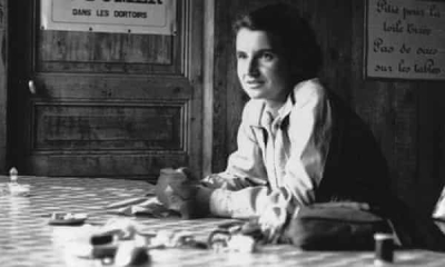
Rosalind Franklin is an English chemist, best known for her role in the discovery of the structure of DNA, a constituent of chromosomes that serves to encode genetic information. Her work on the X-ray diffraction images of DNA, particularly Photo 51, led to the discovery of the double helix shape of DNA. Since Nobel Committee does not recognise work posthumously, the Nobel Prize in Physiology or Medicine in 1962 went to Francis Crick, James Watson, and Maurice Wilkins, who based their work on her data.

Rosalind was a topper and an all-rounder in school. Her interests were in maths, sports and languages. Born to a prominent British Jewish family in 1920, Franklin studied the Natural Sciences Tripos at Newnham College, Cambridge, from which she graduated in 1941. She joined the University of Cambridge physical chemistry laboratory as a research fellow. Since this was during World War II, she worked on the porousity of coal for fuel purposes and other wartime devices

After finishing her work on DNA, Franklin led pioneering work at Birkbeck, University of London, on the molecular structures of the Tobacco Mosaic virus (TMV), an RNA virus that infects tobacco plants. Her work provided new insights into the structure of viruses.

Picture Credit : Google

What is Pasteurisation technique of Louis Pasteur?

Pasteur’s experiments with germs and wine revealed a direct cause-and-effect relationship between bacteria and the souring of wine into vinegar. Subsequently, he invented a process by which bacteria could be killed by heating the wine between 60 and 100° C, then letting it cool. Pasteur completed the first successful test on April 20, 1862, eventually patenting the method we now know as pasteurization, which was soon applied to beer, juice, eggs, and (most famously) milk. This process also proved successful at destroying most yeasts and molds without causing a phase transition in the product.

The temperature and time of pasteurization treatments are determined by the food’s acidity. In acidic foods (pH < 4.6) such as fruit juice, in which pathogens are unable to grow, heat is applied to inactivate enzymes and destroy yeast and lactobacillus. In less acidic foods (pH > 4.6), such as milk, the heat treatments are designed to destroy pathogens, as well as yeast and molds. Both processes extend the product’s shelf life, especially in combination with refrigeration.

Food can be pasteurized in two basic ways: either before or after being packaged into containers. When food is packaged in glass, hot water is used to lower the risk of thermal shock. If packaged in plastic or metal, steam can be used, since the risk of thermal shock is low. In general, most foods requiring pasteurization are liquid (such as milk), and can therefore move through a continuous system comprised of a heating zone, hold tube, and cooling zone, from which the liquid is filled into packaging.

Credit : Smart Sense

Picture Credit : Google

What are the two diseases that Louis Pasteur developed vaccines for?

His research, which showed that microorganisms cause both fermentation and disease, supported the germ theory of disease at a time when its validity was still being questioned. In his ongoing quest for disease treatments he created the first vaccines for fowl cholera; anthrax, a major livestock disease that in recent times has been used against humans in germ warfare; and the dreaded rabies.

Pasteur produced vaccines from weakened anthrax bacilli that could indeed protect sheep and other animals. In public demonstrations at Pouilly-le-Fort before crowds of observers, twenty-four sheep, one goat, and six cows were subjected to a two-part course of inoculations with the new vaccine, on May 5, 1881, and again on May 17. Meanwhile a control group of twenty-four sheep, one goat, and four cows remained unvaccinated. On May 31 all the animals were inoculated with virulent anthrax bacilli, and two days later, on June 2, the crowd reassembled. Pasteur and his collaborators arrived to great applause. The effects of the vaccine were undeniable: the vaccinated animals were all alive. Of the control animals all the sheep were dead except three wobbly individuals who died by the end of the day, and the four unprotected cows were swollen and feverish. The single goat had expired too.

As with other infectious diseases, rabies could be injected into other species and attenuated. Attenuation of rabies was first achieved in monkeys and later in rabbits. Meeting with success in protecting dogs, even those already bitten by a rabid animal, on July 6, 1885, Pasteur agreed with some reluctance to treat his first human patient, Joseph Meister, a nine-year-old who was otherwise doomed to a near-certain death. Success in this case and thousands of others convinced a grateful public throughout the world to make contributions to the Institut Pasteur. It was officially opened in 1888 and continues as one of the premier institutions of biomedical research in the world. Its tradition of discovering and producing vaccines is carried on today by the pharmaceutical company Sanofi Pasteur.

Credit : Science History

Picture Credit : Google

In which year did Linda Buck receive the Nobel Prize for Medicine?

Linda B. Buck, American scientist and corecipient, with Richard Axel, of the Nobel Prize for Physiology or Medicine in 2004 for discoveries concerning the olfactory system.

In 1991 Buck and Axel jointly published a landmark scientific paper, based on research they had conducted with laboratory rats, that detailed their discovery of the family of 1,000 genes that encode, or produce, an equivalent number of olfactory receptors. These receptors are proteins responsible for detecting the odorant molecules in the air and are located on olfactory receptor cells, which are clustered within a small area in the back of the nasal cavity. The two scientists then clarified how the olfactory system functions by showing that each receptor cell has only one type of odour receptor, which is specialized to recognize a few odours. After odorant molecules bind to receptors, the receptor cells send electrical signals to the olfactory bulb in the brain. The brain combines information from several types of receptors in specific patterns, which are experienced as distinct odours.

Axel and Buck later determined that most of the details they uncovered about the sense of smell are virtually identical in rats, humans, and other animals, although they discovered that humans have only about 350 types of working olfactory receptors, about one-third the number in rats.

Credit : Britannica

Picture Credit : Google

What is Jarvik 7?

You may have heard of heart transplants, where organs of diseased people are transplanted into living recipients. In many such cases there is a long list of recipients and a very short list of donors. But did you know that instead of waiting for a heart donor, there is another option for heart patients? The alternative is called the ‘Total Artificial Heart’ or TAH. The TAH is a form of mechanical circulatory support in which the patient is supported by a pneumatically powered, artificial heart.

The first TAH to be implanted was the Jarvik 7 in 1982. The Jarvik 7 was an artificial heart made of plastic and metal, named after its designer Robert Jarvik. It was the first TAH which was designed to be implanted as a permanent replacement after the natural heart was removed.

In probably one of the most dramatic surgical procedures that was ever imagined, surgeons at the University of Utah implanted the Jarvik 7 heart into Barney Clark, a 62-year-old dentist, in 1982.

Clark, who was in the final stages of heart failure, agreed to the procedure as an experiment to help medicine and did not expect to survive more than a few days. He however, went on to live for 112 days after the implantation.

With the relative success of the first operation, by the late 1980s, surgeons at 16 centers were using the Jarvik 7 as a bridge to heart transplantation. Today, though the original Jarvik 7 heart is no longer in use, this invention was truly a remarkable feat in the history of medical science.

Picture Credit : Google

What is MRI and how is it useful?

Magnetic Resonance Imaging, or MRI, is a valuable clinical and research tool. It gives a visual representation of organs and structures in the body.

Magnetic resonance imaging was made possible when Nikola Tesla first discovered the rotating magnetic field in 1882. Nearly one hundred years later in 1977, an American physician, Raymond Vahan Damadian invented the first MRI scanning machine. He was the first to perform a full body scan on a human being with a resultant diagnosis of cancer.

An MRI scanner consists of a long tube that the patient is inserted into. Here the patient is subjected to a very strong magnetic field and radio waves are applied in different directions. This causes atoms in the patient’s body to emit special signals which are detected by the MRI scanner and sent to a computer. Based on the data the computer creates an image of the part of the body to be scanned.

Although it is more expensive than other procedures, a major advantage of MRI is that dangerous X-ray radiation can be avoided. MRI’s are used to study the brain, spinal cord, bones, joints and internal organs. It is an extremely accurate diagnostic method and an invaluable tool for disease detection.

The development of MRI revolutionized the medical world. Since its discovery, MRI scans not only assist in medical procedures but also aid in research.

Picture Credit : Google

What is a CAT Scan?

A CAT scan or CT scan is a special X-ray test that produces cross-sectional or three-dimensional images of different sections of the body. The acronym ‘CAT’ stands for ‘Computerized Axial Tomography’.

The invention of computers sparked researchers to try to combine X-rays with computer technology. In 1972, a British biomedical engineer named Sir Godfrey Hounsfield, developed the CAT scan. He received the Nobel Prize in 1979 for his work.

The first clinical CT scanners began to be installed in 1974. The original systems were dedicated to taking images of the head only, but ‘whole body’ systems became available in 1976. CT scans became widely available by about 1980.

The first CT scanner took several hours for a single scan and took days to reconstruct a single image from raw data. Today, an entire chest CT of 40 slices and 8mm thickness, can be scanned in 5 to 10 seconds using the most advanced multi-slice CT system.

During its 25-year history, CT has made great improvements in speed, patient comfort and resolution. CT scanners are now used to take pictures of any part of the body and are invaluable to medical diagnosis.

Picture Credit : Google

How has heart transplant surgery changed medical history?

In a heart transplant procedure, a surgeon removes the diseased heart of a person and replaces it with a donor heart. It is used to treat the most serious cases and as an option for people in the final stages of heart failure.

This sort of surgery was unimaginable till the beginning of the 20th century. It was only the invention of the heart-lung bypass machine by John Gibbon, in 1953 that made such a procedure possible. In 1967, for the first time, a human heart from one person was transplanted into another by a surgeon named Christiaan Barnard in Cape Town, South Africa.

Dr Barnard’s team removed the heart of a 25-year-old woman, who had died following an auto accident, and placed it in the chest of Louis Washkansky, a 55-year-old man dying of heart damage. The surgery was the first of its kind and the patient survived for 18 days on the borrowed heart.

In the 1970s, with the development of better anti-rejection drugs, heart transplantation became more viable and by the late 1970s patients were living up to five years with their new hearts. Today, new approaches to heart transplant surgery are being researched, but Barnard’s operation will always be remembered as a historic achievement in the field of medicine.

Picture Credit : Google

What is polio and who was behind the polio vaccine?

Polio is caused by one of three types of poliovirus from the Enterovirus genus. It spreads through contact between people, through nasal and oral secretions and through contaminated faeces. The polio virus can damage the brain and spinal cord. In around 98 per cent of cases it is a mild attack with almost no symptoms. However in a very small percentage of people, it can cause paralysis and loss of the use of a limb.

Polio has been around for thousands of years and affected human populations at relatively low levels till the early 1900s. This was a time when other diseases like diphtheria, typhoid and tuberculosis were being treated successfully. Ironically, advances in hygiene led to the increase of polio cases. This was because in the past, infants were exposed to the polio virus through contaminated water supplies at early ages. These exposed infants developed immunity due to natural maternal antibodies still circulating in their system. However, better sanitation meant that exposure to the polio virus was getting delayed till children had lost their maternal antibodies, which led to them being more vulnerable.

By 1940, polio was a highly infectious disease and many scientists were racing to make a vaccine for it.

It was Dr Jonas Salk who finally succeeded in 1955. He started working on a vaccine against the three strains of polio and tested the vaccine on his own children first. Salk’s vaccine was composed of an injected, inactivated form of the polio virus, which retained the ability to give Immunity against the disease without the risk of infecting the patient.

However, Salk’s vaccine was not completely effective against one of the three strains of polio and it only gave immunity for a limited time. In 1957, Albert Sabin, a Polish- American medical researcher, succeeded in making an oral vaccine which used a small amount of the live virus. This vaccine became available globally in 1962 and today, the oral polio vaccine is one of the most successful vaccines of all times.

Currently, the polio virus is only found in Afghanistan and Pakistan with an occasional spread to neighbouring countries.

Picture Credit : Google

What is an endoscope and why is it important?

An endoscope is an instrument that allows a doctor to look inside the human body. Evidence of such instruments have been found since early Greek and Roman times. In 1805, Philip Bozzini made the first endoscope called ‘lichtleiter’ which means light guiding instrument. He used this tube to examine the pharynx of a living person for the first time.

In 1853 Antoine Jean Desormeaux first developed an instrument specially designed to examine the urinary tract and the bladder. He was the first to coin the term ‘endoscope’. In 1932, Dr Rudolph Schindler invented a flexible gastroscope and examined the inside of a stomach through numerous lenses positioned throughout the tube. In 1965, Harold Hopkins introduced rod lenses to give a clearer view.

Today’s endoscopes use optical fibres. One of the two main cables of the endoscope carries light down into the body while the surgeon looks down the other which has a camera. This camera sends back information to a computer. The procedure is conducted to look at internal organs and structures and is an important aid to diagnosis.

Picture Credit : Google

Why are tranquillisers considered to be wonder drugs?

Tranquillisers are drugs that are prescribed by a doctor to reduce anxiety, fear, tension and other forms of mental disturbance. An Australian psychiatrist, Dr John Cade, was the first to use a tranquilliser to calm a mentally ill patient. Tranquillisers were first developed in the early 1950s, not as a cure but to relieve symptoms associated with other problems.

They help to calm agitated, excited and irrational patients and enable many seriously ill people, who would otherwise be hospitalized, to live at home, and engage in productive work.

Tranquillisers are also found to be useful during surgery to reduce the amount of anaesthetic needed. Before the advent of tranquillisers, the treatment of mental illness was often cruel and drastic and some of the procedures left patients unfit to lead a normal life. To this end, tranquillisers are indeed wonder drugs.

Picture Credit : Google

Why the discovery of DNA was considered a silent revolution in genetics?

DNA is short for ‘deoxyribonucleic acid’. It is a complex molecule that holds the genetic code (or blueprint) of all organisms including humans, animals and plants. DNA determines, among other things, how tall or short you are, the colour of your skin, or if you have brown eyes or blue.

It is present in each cell of the organism and is inherited from parents to children. The theory of genetic inheritance was first proposed by Gregor Mendel in 1865. He suggested that information was passed on in discrete units from parent to child and that traits were not ‘blended’ together. His findings formed the basis of the science of genetics.

DNA was discovered accidentally in 1869 by Friedrich Miescher who was studying the composition of lymphoid cells (white blood cells). He isolated a new molecule he called nuclein from a cell nucleus. He was the first to define DNA as a distinct entity.

After much research, in 1953 James Watson and Francis Crick presented the double-helix structure of DNA, consisting of two DNA strands wound around each other. It was shown that each of the inter-twined strands of DNA consisted of a chain of chemical groups called nucleotides.

Nucleotides are made up of four chemical bases. The order of these bases determines the information available for building and maintaining an organism.

The discovery of the structure of DNA transformed science, medicine and agriculture. It revolutionized the investigation of disease and the identification of pathogens. It is also a major component of forensic science where evidence based on DNA findings helps solve many cases.

When Watson and Crick discovered the structure of DNA, they had stumbled upon the secret of all life on earth!

Picture Credit : Google

Who developed the kidney dialysis machine?

Kidney failure can lead to death if untreated and is an illness that is as old as humanity itself. In 1924, a German doctor named Georg Haas carried out the first dialysis treatment on a human. However, this treatment was ineffective and the patient did not survive.

It was a Dutch doctor, Willem Kolff, who conducted the first successful dialysis treatment of a 67-year old patient. The patient was discharged with normal kidney function after a week.

Kolff used a rotating drum kidney or artificial kidney to filter uremic toxins from the body. His model improved on the earlier one made by Haas by using membranous tubes made from cellophane. Kolff began work on his artificial kidney in the late 1930’s after watching a young man die of kidney failure. Kolff decided that he would make a machine that would do the work of the kidneys.

Over the years, Kolff improvised - using sausage skins, orange juice cans, a washing machine and other common items to make a device that could clear the blood of toxins. He created the first successful artificial kidney in 1945. One of the drawbacks to Kolff’s machine was that while the Kolff kidney effectively removed toxins from the blood, it was unable to remove excess fluid from the patient’s blood.

Today dialysis machines are designed to filter out excess fluid as well as toxins and are highly sophisticated. However, Kolff’s machine is considered the first modern drum dialyzer and it remained the standard over the next decade.

Picture Credit : Google

When was streptomycin invented?

Streptomycin is an antibiotic used to treat bacterial infections. It was the first antibiotic effective against tuberculosis and was discovered in 1943. 

After the discovery of penicillin, researchers were inspired to search for more antibiotics. At this time, Selman Abraham Waksman, a Jewish-American microbiologist, was researching the decomposition of organisms that lived in the soil. He was working closely with a bacteria called ‘streptomyces griseus’ but was unable to produce consistent cultures.

A breakthrough occurred when a farmer took one of his hens, suffering from a mysterious disease, to the research station at Rutgers University where Waksman worked. A vet took a sample from the hen’s throat, analyzed it and found that it was a strain of streptomyces griseus. It was duly sent to Waksman’s lab for testing. Here, a PhD student named Albert Schatz, isolated a substance from this bacterium, which was found to be consistently effective against tuberculosis. Schatz named the substance ‘streptomycin’.

Streptomycin was also found to be effective against meningitis, typhoid and even the plague! Waksman, who developed a number of antibiotics, was responsible for introducing procedures that led to the development of many more. He was awarded the Nobel Prize for his work in 1952.

Picture Credit : Google

What is medical ultrasound?

UItrasonography or ultrasound is the use of high-frequency sound waves to produce images of structures within the body.

These sound waves are inaudible to humans and are produced by the electrical stimulation of a ‘piezoelectric’ crystal. The waves are aimed at a specific part of the body and are reflected back according to changes in tissue density. The relative position of tissues is gauged by an electronic apparatus which forms images that are displayed as ultrasound scans. These scans provide valuable information for diagnosing and treating a variety of diseases and conditions.

The main advantage of the ultrasound is that sound waves are less harmful than X-rays and can be safely used on pregnant women and children.

Ultrasound technology developed as part of echolocation studies beginning in the late 18th century. Austrian neurologist Karl Dussik was the first person to utilize ultrasound for medical purposes. In 1942, he detected brain tumours in a patient using ultrasound.

Ultrasound was introduced in the field of obstetrics in 1958 by English physician Ian Donald. In the 1980’s the Japanese began experimenting with 3D ultrasound. Rapid technological advances in electronics provided further improvements. Today 4D ultrasounds, in which the ultrasound shows movement, are common.

Picture Credit : Google