Did you know scientists hope to develop targeted drugs for hard-to-treat diseace using the technique?
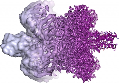
Ultra high-definition 3D videos of cells inside the body taken by an advanced microscope are creating a revolution in modern biology. Previously, transmission electron microscope (TEM) images of living cells (called biomolecules) were indistinct blobs. This was because the high-energy electron beams would dry out the water surrounding the molecules and burn them.
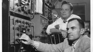
In cryogenic electron microscopy (cryo-EM), biomolecules are cooled to extremely low temperatures and embedded in vitrified water (ice that has no crystals). This ensures that they are preserved intact. The three scientists who developed the cryo-EM technique won the 2017 Nobel Prize in Chemistry.
The current level of sophistication in the cryo-EM technique is largely due to advances in camera technology, image processing and computer software. The biomolecules are photographed from thousands of different angles and at different stages. The images are put together to create a video, allowing researchers to see the structure of the molecules in live action within the cell.
Using this technique, scientists hope to develop targeted drugs for hard-to-treat diseases such as dementia and Parkinson’s.
Picture Credit : Google