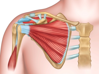
What is the anatomy of shoulder?


As well as controlling a wide range of arm movements, the shoulder muscles also stabilize the joint so that it doesn’t dislocate (pop out of its socket).


Trapezius


This helps to move the shoulder blaze. The trapezius muscle is a large, triangular, paired muscle located on the posterior aspect of the neck and thorax. When viewed together, this pair forms a diamond or trapezoid shape, hence its name.


Clavicle (collarbone)


The clavicle, or collarbone, is a long bone that serves as a strut between the shoulder blade and the sternum (breastbone). There are two clavicles, one on the left and one on the right. The clavicle is the only long bone in the body that lies horizontally. Together with the shoulder blade, it makes up the shoulder girdle.


Subscapularis


Subscapularis is a triangular shoulder muscle located in the subscapular fossa of scapula. Attaching between the scapula and the proximal humerus, it is one of the four muscles of the rotator cuff, along with supraspinatus, infraspinatus and teres minor. The arm can be twisted towards by this muscle.
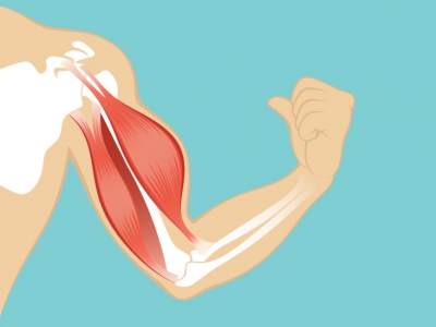


Coracobrachialis


The coracobrachialis is a long and slender muscle of the anterior compartment of the arm. As its name suggests, it extends from the coracoid process of scapula to the shaft of the humerus. This muscle helps to flex the shoulder and pull the upper arm in towards the body.


Humerus


The humerus is the bone of the upper arm. It consists of a proximal end, a shaft and a distal end, all which contain important anatomical landmarks. 


Biceps brachii


The biceps brachii muscle is one of the chief muscles of the arm. This is the muscle that bends the elbow. The origin at the scapula and the insertion into the radius of the biceps brachii means it can act on both the shoulder joint and the elbow joint, which is why this muscle participates in a few movements of the arm.


Picture Credit : Google


What is the function of shoulder?


The shoulder joint – where the upper arm and shoulder blade meet – is the most flexible joint in the body. This mobility, combined with long arms and grasping hands, enables humans to perform a huge range of arm movements.


The bony framework of the shoulder joint is formed by three bones – the shoulder blade (scapula), the collarbone (clavicle), and the top of the upper arm bone (humerus). Deep muscles make the joint stable, while an outer layer of muscles pulls on the bones to move the joint.


The shoulder has about eight muscles that attach to the scapula, humerus, and clavicle. These muscles form the outer shape of the shoulder and underarm. The muscles in the shoulder aid in a wide range of movement and help protect and maintain the main shoulder joint, known as the glenohumeral joint.


Picture Credit : Google


What is the function of arms and hands?


The upper limbs are the body’s most flexible parts. Shoulder and elbow joints allow the arms to move in all directions. Attached to the arms are our versatile hands. They have countless capabilities, allowing us to touch, lift, throw, and grip. A team of muscles, tendons, and ligaments move and support these limbs.


Hand function has great significance for occupational performance. The greater the difficulties with hand function, the greater the impairment in skills that allow for independence and participation in academic and social activities.


Your arms contain many muscles that work together to allow you to perform all sorts of motions and tasks. Each of your arms is composed of your upper arm and forearm. Your upper arm extends from your shoulder to your elbow. Your forearm runs from your elbow to your wrist.


Picture Credit : Google


How speaking works?


As well as supplying the body with oxygen, breathing is also essential for another job; making sounds using the voice. Humans are social beings, so communicating with those around us is very important. We use our voices to deliver information or express our feelings through talking, laughing, or even singing.


How speaking works


As air is breathed out, it passes through the voice box (larynx), below the back of the tongue. Stretched across the larynx are two flexible membranes called the vocal cords. When we want to speak, muscles pull the vocal cords closer together. Air pushes through the small gap, making the cords vibrate to produce a sound. This sound is shaped into a series of words by moving the mouth, lips, and tongue into different positions.


High and low pitch


People’s voices have different tones and pitches. Men tend to have deeper voices, as their vocal cords are long and thick, producing a lower sound. Women have higher-pitched voices, and children’s are the highest of all, because their vocal cords are much shorter.


Range of the human voice


We measure pitch in hertz (Hz), which describes how fast the vocal cords vibrate per sound (frequency).


Picture Credit : Google


What is abnormal breathing called?


Normal breathing happens in a regular, repeated pattern. Sometimes, though, there’s a different kind of breath, such as a cough, sneeze, or hiccup.


Sneezing


When something irritating gets inside the nose, the brain’s response is to trigger a sneeze to clear the air passages. After a sharp breath, muscles in the chest and abdomen contract to force the air back out, carrying the intruding particles with it.


Snoring


Sometimes, the breaths we take while asleep can be heard – in fact, the sound can be loud enough to wake the sleeper. Snoring is caused by relaxed tissues vibrating as air passes over them.


Hiccups


Hiccups happen when nerves around the diaphragm get irritated, for example by eating quickly. The diaphragm jerks, making the lungs suddenly suck in air. This makes the vocal cords snap shut, making a “hic!” sound.


Coughing


Coughing occurs when the body tries to clear something irritating, such as smoke, from the airways. The vocal cords close, so that no air can get though. Then the lungs push air against the cords so they suddenly open, releasing an explosive breath.


Yawning


A yawn is a deep breath with the mouth wide open, stretching the eardrums and muscles around the throat. Though we often yawn when we’re tired, no one knows exactly what yawning is for. It may help to cool the brain, or help to keep you awake and alert.


Picture Credit : Google


What is meant by rate of breathing?


Fuelling the muscles


With each breath we take, oxygen is delivered to the muscle cells to provide the energy that powers movement. The faster the body moves or the harder it works, the more oxygen the cells need. So our breathing speeds up in order to take in, then deliver, more oxygen to where it is needed.


Breathing rates


Breathing rate, or speed, depends on a person’s age, size, health, and fitness level as well as on what they are doing. This shows a typical adult’s breathing rates when they are taking part in different activities.


A normal breathing rate for an adult at rest is 8 to 16 breaths per minute. For an infant, a normal rate is up to 44 breaths per minute.


  • Reading 15 breaths per minute

  • Walking 20 breaths per minute

  • Jogging 40 breaths per minute

  • Running fast up to 70 breaths per minute


Fitness and breathing


When we exercise, we breathe heavily to take in extra oxygen. With regular exercise, the lungs increase their ability to hold air, and our bodies get more efficient at using the oxygen they take in. this means we don’t have to breathe as fast to get the same amount of oxygen to the muscles.


Fighting fit


The more physically fit a person is, the easier they will find it to work out in the gym, dance, or run for a bus, without panting or getting out of breath.


Picture Credit : Google


How breathing works?


When the lungs take a breath in, they expand – but they can’t do this on their own. To make them suck in air, the lungs are pulled open by the muscles around them. Then, to breathe out, the muscles relax and the lungs become smaller again, so air is squeezed back out. The muscles used for breathing are the diaphragm, which is below the lungs, and the intercostals muscles, between the ribs.


Breathe in and out


To breathe in, muscles between the ribs contract, pulling the ribcage upwards and outwards. At the same time, the diaphragm, a strong sheet of muscle below the lungs, tightens, pulling the lungs down. The lungs expand and suck in air.


To breathe out, the rib muscles and diaphragm relax again. This makes the ribcage fall back down, while the diaphragm moves upwards. The lungs are squeezed and become smaller, pushing air back out.


Air enters through mouth and nose. As the ribcage is pulled up and out, the chest expands. The lungs enlarge to fill the larger space in the chest. The diaphragm contracts, pulling the bottom of the lungs down.


Air leaves the body. The falling ribcage pushes the lungs down and in. The rib and chest muscles relax, tilting the ribs downwards. The lungs are made smaller, forcing out air. The diaphragm relaxes and becomes longer and more flexible.


Picture Credit : Google


How does breathing affect speech?


Humans need to breathe almost constantly, in order to provide all the body’s cells with the oxygen they need to keep working. We don’t need to remember top breathe – the brain makes sure we do it automatically, even when we’re asleep. But we can also take control of our breathing, so that we can perform actions such as talking, singing, playing a wind instrument, or just blowing out the candles on a birthday cake. The breath is used for other actions too, such as sneezing and coughing.


There is a difference between breathing for life and breathing for speech. Breathing properly is an important skill for producing a stronger voice with better projection. Without proper breathing, it may be difficult to fully achieve your speech and voice goals.


Many individuals breathe in a way that prevents them from producing a voice that is strong and rich in tone. Speaking with a fuller breath will allow you to feel confident and physically more comfortable, give your message more energy and speak with vibrant vocal tone quality.


Picture Credit : Google


How does gas exchange take place in the lungs?


Every second of every day a life-giving process called gas exchange occurs in the lungs. During gas exchange, oxygen enters the bloodstream and carbon dioxide – a waste product – leaves.


The lining of stretchy air pockets called alveoli. Red blood cells flow through tiny blood vessels that surround the alveoli. The cells pick up oxygen and carry it away in the bloodstream to the body’s tissues and organs. White blood cells work for the immune system by surrounding and destroying bacteria and other germs.


The body's circulation is an essential link between the atmosphere, which contains oxygen, and the cells of the body, which consume oxygen. For example, the delivery of oxygen to the muscle cells throughout the body depends not only on the lungs but also on the ability of the blood to carry oxygen and on the ability of the circulation to transport blood to muscle. In addition, a small fraction of the blood pumped from the heart enters the bronchial arteries and nourishes the airways.


Picture Credit : Google


What is responsible for swapping gases?


Carbon dioxide leaves the blood and passes through the thin walls of the capillary and alveolus into the air, ready to be breathed out. Oxygen moves in the opposite direction, into the blood stream.


To support the absorption of oxygen and release of carbon dioxide, about 5 to 8 liters (about 1.3 to 2.1 gallons) of air per minute are brought in and out of the lungs, and about three tenths of a liter (about three tenths of a quart) of oxygen is transferred from the alveoli to the blood each minute, even when the person is at rest. At the same time, a similar volume of carbon dioxide moves from the blood to the alveoli and is exhaled. During exercise, it is possible to breathe in and out more than 100 liters (about 26 gallons) of air per minute and extract 3 liters (a little less than 1 gallon) of oxygen from this air per minute. The rate at which oxygen is used by the body is one measure of the rate of energy expended by the body. Breathing in and out is accomplished by respiratory muscles.


Three processes are essential for the transfer of oxygen from the outside air to the blood flowing through the lungs: ventilation, diffusion, and perfusion.


Picture Credit : Google


What is the function of lungs?


The two lungs take up most of the space in the chest. Their key function is to get oxygen into, and waste gases out of, the bloodstream. That oxygen is used by the body’s cells to release energy, a process that produces waste carbon dioxide.


Breathing draws air rich in oxygen into the lungs through the airways, then pushes air containing carbon dioxide in the opposite direction. Lungs are spongy because they are packed with branching, air-filled tubes that get narrower and narrower before ending in tiny air sacs (alveoli). It is here that oxygen is swapped for carbon dioxide.


The lungs are like bellows. As they expand, air is sucked in for oxygen. As they compress, the exchanged carbon dioxide waste is pushed back out during exhalation.


When air enters the nose or mouth, it travels down the trachea, also called the windpipe. After this, it reaches a section called the carina. At the carina, the windpipe splits into two, creating two mainstem bronchi. One leads to the left lung and the other to the right lung.


From there, like branches on a tree, the pipe-like bronchi split again into smaller bronchi and then even smaller bronchioles. This ever-decreasing pipework eventually terminates in the alveoli, which are little air sac endings.


Picture Credit : Google


How blood clots?


After a cut, blood seeps from the wound, triggering an immediate repair process. The blood cells take action immediately. They stop the leak, form a plug, and destroy harmful bacteria. A scab forms, and the clot dissolves when the wound has healed.


Injury


A cut in the skin damages blood vessels. Platelets start to group together at the site of the injury.


Plug


The platelets release chemicals that make fibrin, a sticky thread-like protein. Red cells get stuck in the threads, forming a plug. White blood cells arrive to hunt for germs.


Clot


The fibrin threads contract, binding red blood cells and platelets together in a sticky clot, which closes the wound.


Scab


The clot near the skin’s surface dries out to form a protective scab, which covers the healing wound.


Picture Credit : Google