What is in the female reproductive system?


Inside the reproductive system


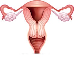
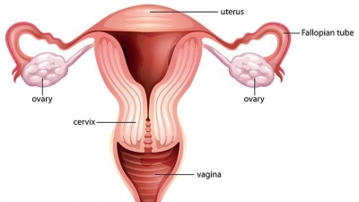
The uterus is in the middle of the lower abdomen, between the bladder and the rectum. The two ovaries are on either side of the uterus, connected to it by the fallopian tubes.


Inside the ovary


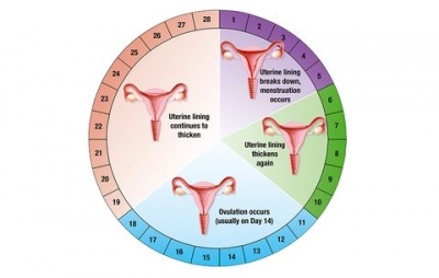
The ovaries contain many thousands of immature eggs, each enclosed in a bag-like follicle. Every month, hormones trigger a process where one of the eggs starts to outgrow the others. When it is mature, the egg is released from the ovary.


Right ovary


The ovary is a ductless reproductive gland in which the female reproductive cells are produced. Females have a pair of ovaries, held by a membrane beside the uterus on each side of the lower abdomen. The ovaries store, then release eggs.


Right fallopian tube


The uterine tubes, also known as oviducts or fallopian tubes, are the female structures that transport the ova from the ovary to the uterus each month. Each fallopian tube connects an ovary with the uterus.


Uterus


This hollow, stretchy organ is where the embryo develops. It functions to nourish and house a fertilized egg until the fetus, or offspring, is ready to be delivered.


Cervix


The cervix is the lower portion of the uterus, an organ of the female reproductive tract. The cervix is the point where the uterus opens into the vagina. Anatomically and histologically, the cervix is distinct from the uterus, and hence we consider it as a separate anatomical structure.


Bladder


This stretchy bag stores and releases urine. The bladder is a pyramidal shaped organ, which sits in our pelvis (the bony structure which helps form our hips).

The main function of the bladder is to store urine, and under the appropriate signals, release it into a tube which carries the urine out of the body.


Rectum


The lowest part of the large intestine is where faeces are stored. The rectum begins at the height of S2-S3 and ends at the perineum. It is about 12 to 16 cm long.


Vagina


The vagina is a fibromuscular (made up of fibrous and muscular tissue) canal leading from the outside of the body to the cervix of the uterus or womb. It is also referred to as the birth canal in the context of pregnancy. The uterus is linked to the outside of the body by this stretchy tube.


Pelvic floor


The uterus and bladder are supported by these strong muscles. These muscles extend across the pelvic region, below other muscles that give humans the ability to walk upright.


Picture Credit : Google


What are the phases of Menstrual Cycle?


Every month or so, a woman’s body goes through the process of preparing for a possible pregnancy. This sequence is called the menstrual cycle.


Phase 1


Egg ripens: Prompted by hormones, an immature egg starts to grow inside a soft casing called a follicle. Your period may last 2 – 7 days, with most fluid being lost in the first two days.


Phase 2


Egg is released: The uterus lining thickens to prepare to receive an egg. Meanwhile, the follicle bursts and releases a ripe egg into the fallopian tube.  During this phase, due to the oestrogen, your skin could be smoother, your breasts a little bigger and any discharge will be clear.


Menstrual cycle: This shows a typical 28-day menstrual cycle. In fact, it can take from 21 to 35 days to complete, and the times between stages vary a good deal.  


Phase 3


Egg arrives in the uterus: At this point, if the egg is fertilized, it embeds itself in the uterus wall and starts to grow. You may feel slightly ‘bloated’ as this phase can trigger fluid retention throughout your body – but it’s not dangerous and doesn’t last.


Phase 4


Menstruation: If the egg is not fertilized, both the egg and the uterus lining fall apart and leave the body along with some blood. This is called menstruation, or having a period. This sudden change can cause mood swings (also called PMS Pre-Menstrual Stress) and you may also experience cramps as the uterus contracts in readiness to shed the built-up layer – which takes you back to Phase 1; having your period.


Picture Credit : Google


What is female reproduction?


From her teenage years to about her mid-fifties, the role of a woman’s reproductive organs is, combination with a man’s sex cells (sperm), to create a new human life – a baby.


A woman’s main reproductive organs are her ovaries and uterus. The two ovaries are where eggs are stored, then released at regular intervals. If an egg is fertilized by male sperm, the job of the uterus is to nurture and protect the egg as it develops – first into an embryo, then a foetus, which grows into a baby, ready to be born.


The vast majority of the eggs within the ovaries steadily die, until they are depleted at menopause. At birth, there are approximately 1 million to 2 million eggs; by the time of puberty, only about 300,000 remain. Of these, only about 500 will be ovulated during a woman's reproductive lifetime. Any remaining eggs gradually die out at menopause.


Picture Credit : Google


How much salt is in a human body?


The water inside our bodies is salty – in fact, it is as salty as seawater. Salt, or sodium, help maintain the body’s water balance – the amount of salt dissolved in the blood tells the hypothalamus how much water the kidneys should release as urine, and how much to keep. Alongside potassium, salt also plays an essential role in helping nerve cells make signals.


The human body contains many salts, of which sodium chloride (AKA common table salt) is the major one, making up around 0.4 per cent of the body’s weight at a concentration pretty well equivalent to that in seawater. A child’s body contains the equivalent of 28 teaspoons of salt, while an adult’s body has 40 teaspoons.


So a 50kg person would contain around 200g of sodium chloride – around 40 teaspoons. Since we lose salt whenever we sweat, it has to be continually replaced.


While excessive consumption of it has been linked to high blood pressure and stomach cancer, salt is a vital part of body chemistry, playing a key role in many functions, from the quality of blood to transmission of nerve signals.


Picture Credit : Google


How does the hypothalamus detect water levels?


The hypothalamus in the brain is responsible for monitoring water level. If it detects too little or too much water, it responds by telling the pituitary gland to release hormones that communicate with me kidneys and other organs.


Too little water


A shortage of water in the body is called dehydration. The body needs to take in more water and also to conserve the water already inside it.


  • Low water alert: The pituitary gland releases a hormone into the bloodstream.

  • Feeling thirsty: The hormone triggers an urge to drink.

  • Dry mouth: The mouth feels dry, as water is sent to areas that need it more.

  • Kidneys: The kidneys receive instructions to remove less water from the blood, so the body produces less urine.


Too much water


Too much water in the body is called overhydration. This condition is rare, but can be caused by illness or by drinking a large amount very quickly. Cells become too waterlogged to work and the blood pressure becomes too high.


  • High water alert: The hypothalamus orders blood vessels to widen, which reduces blood pressure.

  • Kidneys: The kidneys are ordered to extract more water from the blood, making more urine.


Picture Credit : Google


What is in urine test?


Urine can provide valuable clues about health. Dark urine is a sign that a person is dehydrated and needs to drink more. Tests can also detect pregnancy, some infections, hormone changes, and diabetes.


To test urine, a testing strip is dipped into a sample. The coloured bands react to different chemicals in the urine, revealing any abnormalities.


Urine is 94 per cent water. The rest is made up of dissolved substances the body has no use for. They include sodium, which is excess salt, and urea, the waste produced by the liver.


At the microscopic level, your body is constantly working to keep you healthy, even while you sleep. Complex chemical processes take place throughout the body, including the breakdown of proteins known as amino acids. When your body breaks down amino acids, ammonia is left over as waste. That's not something you want in your body for long—ammonia is toxic to human cells.


Since ammonia is toxic to your body, you need a way to remove it. That happens partly in the liver, where the ammonia is broken down into the less-toxic chemical, urea. Urea then combines with water and gets flushed into your bladder through the kidneys as urine, protecting your body from its own chemical processes.


Picture Credit : Google


What are the functions of water in our body?


Water does many different jobs. It helps provide a transport system around the body. It also regulates body temperature, and lubricates parts so they work better.


Blood


Blood is mostly water, so it flows easily through blood vessels. Your blood is 92% water. Water carries nutrients and oxygen to all cells in the body.Water is required for breathing.Water moistens oxygen for breathing


Lymph


Watery lymph flows around the body, recycling chemicals and fighting germs. The lymphatic system works to keep body fluid levels in balance and to defend the body against infections. It is made up of a network of lymphatic vessels that carry lymph — a clear, watery fluid that contains proteins, salts, and other substances — throughout the body.


Saliva


Saliva moistens foods to help with eating and also kills germs in the mouth. Saliva is 99% water and 1% protein and salts. The normal daily production of saliva varies between 0.5 and 1.5 liters. The whole unstimulated saliva flow rate is approximately 0.3-0.4 ml / min. 


Sweat


Sweat is released through skin pores to help cool the body down. Composed of about 99 percent water, sweat evaporates on the skin’s surface, cooling the body and keeping it from overheating.Sweat secretions help you in other ways, too. They include dermcidin, an antibiotic peptide that appears to regulate bacteria growth on the skin and may fight infection.


Urine


Urine is a mix of excess water and chemicals from the blood. This colorless urine is sometimes due to drinking an excess of water, while other times it can signal a problem with the kidneys. If your urine is consistently clear or absent of color, you should see a doctor.


Joints


Many moving joints have a lubricating layer of liquid, called synovial fluid. With its egg white–like consistency, the principle role of synovial fluid is to reduce friction between the articular cartilage of synovial joints during movement. Synovial fluid is a small component of the transcellular fluid component of extracellular fluid.


Tissue fluids


Body tissues contain water, with lean tissue holding more than fatty tissue. Water is also contained inside organs, in gastrointestinal, cerebrospinal, peritoneal, and ocular fluids. Adipose tissue contains about 10% of water, while muscle tissue contains about 75%.


Cell cytoplasm


Cells need water for the chemical reactions that take place inside them. Water is also in the environment outside the cell. For example, water inside cells makes up about 46 percent of your body’s mass, and water outside the cells in body fluids accounts for another 23 percent.


Picture Credit : Google


What is the average percentage of water in the human body?


The amount of water in a person depends on their age, gender and weight. The more water-rich muscles they have, the higher their water content. As we grow older, muscles shrink and water levels drops.


A newborn baby is almost three-quarters water – and the proportion of water in the body drops gradually from then on. Men contain more water than women, as they usually have more water-containing muscle.


The average adult male is about 60% water. The average adult woman is about 55% water because women naturally have more fatty tissue than men. Overweight men and women have more water, as a percent, than their leaner counterparts.


The percent of water depends on your hydration level. People feel thirsty when they have already lost around 2-3% of their body's water. Mental performance and physical coordination start to become impaired before thirst kicks in, typically around 1% dehydration.


Although liquid water is the most abundant molecule in the body, additional water is found in hydrated compounds. About 30-40% of the weight of the human body is the skeleton, but when the bound water is removed, either by chemical desiccation or heat, half the weight is lost.


Body chemistry


Water is essential ingredient of body cells. The millions of chemical reactions that power life take place in the water contained in the body’s cells. Different body tissues contain varying amounts of water, depending on their function. Muscle contains three times more water than bone.


Picture Credit : Google


What are baby turtles called?


A baby turtle is called a hatchling, while hatchling is most often used for a young bird, tadpoles, newts and new born baby turtles.


Baby turtles are cute little creatures that require totally different care than adult turtles, needing a lot of special care to keep them alive and healthy. They're prone to many diseases and carry many disease-carrying germs. You have to feed a baby turtle food that is available in the pet store, while supplementing their diets with vegetables, meat and fruit. Land turtles tend to eat more vegetables than those living in water.


The building block of your baby turtle's diet is the pellet or gel capsule supplement, which is a snap to find in any pet store that sells turtle supplies. There are two caveats, however: you should examine each label to make sure your newborn turtle is getting the best diet possible, and you should select a formula that's been approved for your particular shelled pet. For example, what a baby box turtle eats is more of a true omnivorous diet than a water turtle eats, which includes more raw meat as a baby but a more omnivorous diet as an adult.


Picture Credit : Google


Are turtles reptiles or mammals?


Turtles are reptiles of the order Testudines. They are characterized by a special bony or cartilaginous shell developed from their ribs that acts as a shield. "Turtle" may refer to the order as a whole (American English) or to fresh-water and sea-dwelling Testudines (British English). Testudines includes both extant (living) and extinct species. Its earliest known members date from the Middle Jurassic. Turtles are one of the oldest reptile groups, more ancient than snakes or crocodilians. Of the 356 known extant species, some are highly endangered.


A turtle's diet varies greatly depending on the environment in which it lives. Adult turtles typically eat aquatic plants,[citation needed] invertebrates such as insects, snails, and worms, and have been reported to occasionally eat dead marine animals. Several small freshwater species are carnivorous, eating small fish and a wide range of aquatic life. However, protein is essential to turtle growth and juvenile turtles are purely carnivorous.


Sea turtles typically feed on jellyfish, sponges, and other soft-bodied organisms. Some species with stronger jaws have been observed to eat shellfish, while others, such as the green sea turtle, do not eat meat at all and, instead, have a diet largely made up of algae.


Picture Credit : Google


Can sea turtles feel it when you touch their shell?


Yes, sea turtles can feel it when you touch their shell. Sea turtle shells consist of bones, which are covered by a layer of so-called scutes (plates). These scutes are made of keratin, the same material that human fingernails are made of. There are nerve endings enervating even the bones of the shell. These nerve endings are sensitive to pressure, for example from a touch on the back.


A tortoise’s shell obviously isn’t as sensitive as its skin, but the animals are able to feel the tactile sensations that occur when someone pats, rubs, scratches, or taps it. This has to do with how the shell is physically attached to the tortoise’s body.


One thing that tortoises and turtles have in common is their tendency to recede into their shells when they feel threatened. In the proper care, however, most tortoises appear to enjoy being touched by their caretakers. In some cases, they extend their necks out while being touched or massaged – a sign that the animal wants to be rubbed some more.


Picture Credit : Google


Which part of the brain connects the cerebrum and cerebellum to the spinal cord?


Brainstem: acts as a relay center connecting the cerebrum and cerebellum to the spinal cord. It performs many automatic functions such as breathing, heart rate, body temperature, wake and sleep cycles, digestion, sneezing, coughing, vomiting, and swallowing.


The brain stem has 3 areas:


  • midbrain (also called the mesencephalon)

  • pons

  • medulla oblongata


The brain stem sends information to and from the other parts of the brain to the rest of the body and controls:


  • breathing

  • body temperature

  • blood pressure

  • heart rate

  • hunger and thirst

  • digestion of food


More specific functions of each part of the brainstem are dependent of the cranial nerve nuclei and tracts that reside in each area. The medulla oblongata controls the respiratory function, cardiovascular system, as well as gastrointestinal and digestive activities. The pons is involved in controlling movements of the body and equilibrium. Finally, the midbrain controls eye movements and integrates it with auditory input. 


Picture Credit : Google


What happens after a stroke in the cerebellum?


Before we discuss the various cerebellar stroke effects, it’s important to understand the function and structure of the cerebellum itself. The cerebellum — which in Latin means “little brain” — is the second largest part of the brain, following the cerebrum. The cerebellum is part of the hindbrain, located along the rear base of the skull and tucked just beneath the cerebrum. Although the cerebellum only accounts for roughly 10 percent of the brain’s volume, it contains more than half of its total neurons. This mighty “little brain” is responsible for controlling and regulating balance and coordination. The cerebellum plays a major role in fine motor movements and especially muscle memory. In other words, it controls the movements we learn over time through repetition, such as playing an instrument or throwing a baseball.


After a stroke occurs, immediate medical attention is vital to minimizing neural damage. Signs to look for to determine if a patient has suffered a cerebellar stroke include headache, nausea, loss of coordination, vomiting, and difficulty swallowing. Dizziness is one of the most commonly reported cerebellar stroke symptoms, with nearly three-quarters of stroke patients experiencing instances of vertigo, the sensation of falling to the left or right. Other cerebellar stroke effects such as double-vision or nystagmus (condition involving involuntary eye movements) can exacerbate instances of vertigo. Nausea, with or without vomiting, is also extremely common, affecting more than half of cerebellar stroke patients.


Picture Credit : Google