What is the function of pelvis?


The pelvis is a large bowl-shaped group of bones at the base of the abdomen. This section of the skeleton is made up of several bones fused and linked together. It surrounds and protects the soft organs inside the lower abdomen. The pelvis has many functions. It supports the intestines and bladder and the space in the middle allows waste from the intestines and bladder to leave the body In women, the pelvis supports the uterus as it expands to hold a growing baby and also provides the baby's route out of the body during childbirth. Many muscles in the back, abdomen, and legs are anchored to the pelvis, helping to keep the body upright. The pelvis allows us to stand walk, and run without falling over.


The muscles of the pelvic floor wrap around and control the opening of your bladder and rectum. When there is an increase in abdominal pressure (for example when you cough, sneeze, laugh or jump), these muscles contract around your urethra and anus to prevent leakage. Equally as important, these muscles have to relax and lengthen to allow us to urinate or have bowel movements easily.  


Picture Credit : Google


Does the large intestine digest Chyme?


After hours of mechanical and chemical digestion, food has been reduced into chyme. As particles of food become small enough, they are passed out of the stomach at regular intervals into the small intestine, which stimulates the pancreas to release fluid containing a high concentration of bicarbonate. This fluid neutralizes the gastric juices, which can damage the lining of the intestine and result in duodenal ulcer. Other secretions from the pancreas, gallbladder, liver, and glands in the intestinal wall help in digestion.


When food particles are sufficiently reduced in size and composition, they are absorbed by the intestinal wall and transported to the bloodstream. Some food material is passed from the small intestine to the large intestine. In the large intestine, bacteria break down any proteins and starches in chyme that were not digested fully in the small intestine.


When all of the nutrients have been absorbed from chyme, the remaining waste material changes into semisolids that are called feces. The feces pass to the rectum, to be stored until ready to be discharged from the body during defecation.


Picture Credit : Google


What happens to the food in the large intestine?


At every stage of digestion, food is pushed through the system by a powerful muscle action called peristalsis. The muscles lining the intestine contract then relax in a rhythmic, wave-like action. As the intestine walls are squeezed together, the food is forced further along the tube.


The job of your large intestine is to absorb water, minerals, and some of the remaining nutrients from your food. It will change the leftover waste into a bowel movement. This is also called stool. Your rectum stores the stool until you feel the need to have a bowel movement. Muscles of your rectum then push the stool through your anus and out of your body.


 It takes about 36 hours for food to move through the entire colon. All in all, the whole process — from the time you swallow food to the time it leaves your body as feces — takes about two to five days, depending on the individual.


Picture Credit : Google


What is the anatomy of large intestine?


Up, across, and down


The large intestine is a wide tube that goes around the small intestine. The tube goes up. across the abdomen, then down again. It has a lumpy appearance because of the way the muscles in its wall contract.


Transverse colon


Passing just below the stomach, this is the middle part of the large intestine.  It extends between the right and left colic (splenic) flexures, spanning the right hypochondriac, epigastric and left hypochondriac regions of the abdomen. 


Ascending colon


On the right side of the abdomen, this section of the intestine rises from the caecum. The ascending colon is retroperitoneal and it is connected to the posterior abdominal wall by the Toldt’s fascia. 


Descending colon


The descending colon extends between the left colic flexure and sigmoid colon. It travels through the left hypochondriac region, left flank and left iliac fossa. This section passes down the left side of the abdomen.


lleocaecal valve


A valve stops waste flowing back into the small intestine. An ileocecal valve regulates the passage of intestinal contents from the small into the large intestine. 


Caecum


The caecum receives chyme from the small intestine. The cecum is intraperitoneal with various folds and pockets (retrocecal peritoneal recesses) surrounding it.


Appendix


The vermiform appendix is a blind lymphoid pouch located in the right iliac fossa which arises from the cecum. These two parts of the large intestine are connected by the meso-appendix. This small tube may help digestion by storing "friendly" gut bacteria.


Sigmoid colon


The S-shaped sigmoid colon travels from the left iliac fossa until the third sacral vertebra (rectosigmoid junction). The sigmoid colon contracts forcefully pushing the faeces (waste) into the rectum


Rectum


The rectum is the last section of the large intestine. The typical characteristics of the large intestine (taenia coli, haustra, epiploic appendages) change or even terminate at the rectum. The roles of the rectum include temporary storage of fecal matter and defecation.


Anus


This is where waste leaves the body as faeces. The anal canal forms the terminal part of the gastrointestinal tract. It extends from the anorectal junction to the anus.


Gut bacteria


Trillions of bacteria live in the large intestine. Most are either harmless or actively help to complete digestion by processing the remaining nutrients that could not be digested by enzymes. However, some microorganisms that enter the digestive system can cause illness.


Picture Credit : Google


What is the function of large intestine?


The large intestine is the final stage of the digestive system. It's where most of the water, and the last few nutrients, are taken from the chyme that enters from the small intestine.


It then moves unusable waste out of the body. Most nutrients have been taken from the food before it gets to the large intestine - but there is still vital work for it to do Here, trillions of bacteria help to break the remaining food down into valuable nutrients. The large intestine is wider than the small intestine but it's not nearly as long.


The large intestine has 3 primary functions: absorbing water and electrolytes, producing and absorbing vitamins, and forming and propelling feces toward the rectum for elimination. By the time indigestible materials have reached the colon, most nutrients and up to 90% of the water has been absorbed by the small intestine. The role of the ascending colon is to absorb the remaining water and other key nutrients from the indigestible material, solidifying it to form stool. The descending colon stores feces that will eventually be emptied into the rectum. The sigmoid colon contracts to increase the pressure inside the colon, causing the stool to move into the rectum. The rectum holds the feces awaiting elimination by defecation.


Picture Credit : Google


What is villi?


This magnified image of a scan shows a cut away in the deeply folded lining of the duodenum, the first section of the small intestine. The folds are called villi, and these greatly increase the area of the lining - creating a larger surface through which food nutrients can be absorbed.


Nutrients from food are absorbed by microvilli on the surface of the villi, shown here as a green, fur-like layer. Nutrients then pass through a layer of cells (shown in blue-green) before being carried away by blood vessels in in the middle.


Their function is to increase the surface area of the small intestinal wall for absorption of the digested food. These projections absorb the protein molecules and help in the transfer of the proteins to all cells and tissues. Many blood vessels are present within these villi, that help in the absorption of digested food and carry it to the bloodstream. Later, from the bloodstream, the absorbed food is delivered to each and every cell of the body.


Picture Credit : Google


What is the anatomy of small intestine?


Bundled up


The small intestine is at the front of the lower abdomen, surrounded by the large intestine and other organs. Although it's very long - more than 6 m (20 ft) - the small intestine fits into this space because it is bundled up in a series of loops and coils.


Duodenum


The first part of the small intestine is where bile and enzymes are added to the chyme to help break it down. It extends from the pyloric sphincter of the stomach, wraps around the head of the pancreas in a C-shape and ends at duodenojejunal flexure. The duodenum has four parts: superior (duodenal bulb/ampulla), descending, horizontal and ascending. 


Jejunum


This is the middle section of the small intestine, where most of the digestion and absorption of food takes place. The jejunum is entirely intraperitoneal as the mesentery proper attaches it to the posterior abdominal wall.


Ileum


The final, longest section of the small intestine absorbs some nutrients. It is found in the lower right quadrant of the abdomen, although the terminal ileum can extend into the pelvic cavity. The ileum terminates at the ileal orifice (ileocecal junction) where the cecum of the large intestine begins.


lleocaecal valve


The ileocecal valve is a sphincter muscle situated at the junction of the ileum (last portion of your small intestine) and the colon (first portion of your large intestine). Its function is to allow digested food materials to pass from the small intestine into your large intestine. Chyme from the small intestine passes through here to the large intestine.


Looking inside


A cross-section through part of the small intestine shows the muscles that help to push food along its length. The lining is covered with millions of tiny finger-like projections called villi (a single projection is a villus).


Picture Credit : Google


What is the function of small intestine?


The small intestine is the longest part of the digestive system. It's where most of the digestive process takes place, releasing the nutrients in food so that they can be used to fuel the body's cells.


By the time food reaches the small intestine, the stomach has turned it into a liquid called chyme. This chyme is squirted into the duodenum, the first part of the small intestine, along with bile from the gallbladder and enzymes from the pancreas, which break the chyme down even more. Finally, when most of the food has been broken down into simple nutrients, these pass through the walls of the small intestine and into the bloodstream. The remaining food progresses to the next stage - the large intestine.


As a person grows the small intestine increases 20 times in length from about 200 cm in a newborn to almost 6 m in an adult. The length of the small intestine is approximated by three times the length of the infant, or height of the child or adult.


The duodenum is about 25 cm (10 inches) long; the jejunum is about 2.5 m (8 feet) long and the ileum is about 3.6 m (12 feet) long.


Picture Credit : Google


What is ligament?


Ligaments are made out of connective tissue that has a lot of strong collagen fibers in it. They are found in different shapes and sizes in the body. Some look like pieces of string, others look like narrow or wide bands. There are arch-shaped ligaments, too.


Ligaments often connect two bones together, particularly in the joints: Like strong, firmly attached straps or ropes, they stabilize the joint or hold the ends of two bones together. This ensures that the bones in the joint don’t twist too much or move too far apart and become dislocated.


But there are also some ligaments that aren’t connected to bones. For instance, some make sure that internal organs are kept in place. A typical example is the womb, which is kept in the right position in the pelvis by ligaments. Ligaments may also connect two or more organs to each other. For instance, the liver, intestine and stomach are held in place by ligaments in the abdominal cavity. These ligaments often have sensitive structures like blood vessels or gland ducts running through them. The strong connective tissue in the ligaments protects these structures and prevents them from bending, twisting or tearing.


Picture Credit : Google


Where are Ossicles found?


The ossicles are situated in the middle ear and suspended by ligaments. They articulate with each other through synovial joints to form a chain across the length of the middle ear from the tympanic membrane (laterally) to the oval window (medially). The ossicles transmit mechanical vibrations of the tympanic membrane across this chain to the oval window where fluids of the inner ear will move and excite receptors. This process allows sound to be transformed into electrical signals which are then sent to the brain.  This article will explore the function of the auditory ossicles, their bony features, articulations, associated muscles, and some clinical aspects.


Their role is to mechanically amplify the vibrations of the tympanic membrane and transmit them to the cochlea where they can be interpreted as sound. They are located in the middle ear cavity and articulate with each other via two tiny synovial joints. The stapes also articulates with the oval window via the stapediovestibular joint, which is a syndesmosis 3; this joint transmits the ossicular vibrations to the endolymph in the vestibule.


Interestingly, they are the only bones in the body that do not grow after birth, and are also the smallest bones in the body (variant tiny sesamoids aside).


Picture Credit : Google


What is bone marrow?


Bone marrow is the spongy tissue inside some of the bones in the body, including the hip and thigh bones. Bone marrow contains immature cells, called stem cells.


Most red blood cells, platelets, and most of the white blood cells are formed in the red marrow. Yellow bone marrow produces fat, cartilage, and bone.


White blood cells survive from a few hours to a few days, platelets for about 10 days, and red blood cells for about 120 days. These cells must be constantly replaced by the bone marrow, as each blood cell has a set life expectancy.


Certain conditions may trigger additional production of blood cells. This may happen when the oxygen content of body tissues is low, if there is loss of blood or anemia, or if the number of red blood cells decreases. If these happen, the kidneys produce and release erythropoietin, a hormone that stimulates the bone marrow to produce more red blood cells.


The bone marrow also produces and releases more white blood cells in response to infections, and more platelets in response to bleeding. If a person experiences serious blood loss, yellow bone marrow can be activated and transformed into red bone marrow.


Healthy bone marrow is important for a range of systems and activities.


Picture Credit : Google


Which is the longest and strongest bone in the human body?


The femur bone is the strongest and longest bone in the body, occupying the space of the lower limb, between the hip and knee joints.


The femur is an integral component of ambulation. A lot of the large thigh muscles arise from and insert on the various parts of the femur. Muscles that originate from the pelvis and insert on the anterior or posterior surface of the femur to facilitate flexion and extension around the hips. Muscles which arise from the femur will cross the knee joint to insert on the proximal tibia promote flexion and extension around the knee. The tables below summarize the thigh muscles and their points of origin or insertion with respect to the femur.


Important features of this bone include the head, medial and lateral condyles, patellar surface, medial and lateral epicondyles, and greater and lesser trochanters. The head is where the bone forms the hip joint with the innominate bone. The condyles are the points of articulation (connection) with the tibia, which is a lower leg bone. The patellar surface is the groove where the bone adjoins with the patella, or kneecap. The epicondyles and trochanters are all important attachment sites for various muscles.


Picture Credit : Google


What is Hyoid?


The hyoid bone is a ‘U’ shaped structure located in the anterior neck. It lies at the base of the mandible (approximately C3), where it acts as a site of attachment for the anterior neck muscles.


The hyoid consists of a body, two greater horns, and two lesser horns. The body forms the central quadrilateral-shaped broad segment of the hyoid. The greater horns are larger and longer than the lesser horns of the hyoid. The greater horns and lesser horns are also known as cornu majus and cornu minus, respectively. The body and the greater horns appear to give the hyoid its U-shape with the greater horns forming the limbs of the "U" on either side of the body. The greater and lesser horns normally unite to the body of the hyoid via fibrous tissue or a true joint. As age progresses, there is a physiological progression of ankylosis of the joints connecting the greater and lesser horns with the body of the hyoid.  


The hyoid takes part in all possible functional actions of the orofacial complex. It preserves the patency of the airway between the oropharynx above and tracheal rings below. It also connects to the larynx and hence plays a role in phonation. Other functions include tongue movement, mastication, swallowing, prevention of regurgitation, and even respiration. Furthermore, the hyoid maintains the posture of the head, due to the complex connection it presumes between the mandible and the cervical spine.


Picture Credit : Google
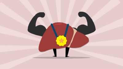

What functions does liver perform?


The liver performs hundreds of essential processing, manufacturing and recycling tasks in the body.


Breaking down


The liver breaks down substances into the parts the body can use or get rid of, such as:


  • Chemicals from food

  • Medicines

  • Germs entering in food


Recycling


It also breaks up dead blood cells so their ingredients can be used again.


Building up


Nutrients are used to make new substances that the body needs, such as:


  • Proteins for building body parts

  • Chemicals to heal injuries

  • Bile, which helps to digest fat


Storage


Useful body substances are stored, then released when necessary, such as:


  • Glucose for energy

  • Minerals, such as iron and copper

  • Vitamins A, D, K and B12


Heating


The liver even gives out heat to help warm the body.


Picture Credit : Google


What is inside the liver?


The liver is in the upper right abdomen, just below the diaphragm. It is divided into two lobes, or sections. An average adult liver is the size of a football and weighs 1.5 kg (3 lb).


Inferior vena cava


It is located at the posterior abdominal wall on the right side of the aorta. The IVC’s function is to carry the venous blood from the lower limbs and abdominopelvic region to the heart. This vein carries blood back to the heart.


Hepatic vein


Oxygen poor blood is drained from the liver to the inferior vena cava in this vein. The main hepatic veins are the right, intermediate and left hepatic veins. In addition, several smaller and somewhat inconsistent caudate lobe veins contribute to the venous drainage of the liver. 


Hepatic duct


The common hepatic duct is the part of the biliary tract formed by the convergence of the right hepatic duct (which drains bile from the right functional lobe of the liver) and the left hepatic duct (which drains bile from the left functional lobe of the liver). This tube drains bile from the right lobe.


Tiny factories


The liver is made up of hexagonal (six-sided) units, called lobules. Each one is the size of a grain of sand. Blood flows from vessels in the lobule corners, is processed by the lobule’s cells, then is collected by the central vein and returned to the heart.


Left lobe


This is the smaller of the two liver lobes. When looking at the front of the liver, the left lobe of liver is divided from the right by the falciform ligament, which attaches the liver to the front wall of the body. The ligamentum venosum and ligamentum teres divide the left lobe of liver from the right as viewed from behind.


Right lobe


This is the biggest section of the liver. The right lobe liver has four sections. It is divided into the anterior right lobe and posterior right lobe by the right hepatic vein. It is also divided into the upper right lobe and lower right lobe by the portal vein.


Ligament


This connective tissue lies between the two main lobes. Liver ligaments are double-layered folds of peritoneum that attach the liver to surrounding organs, or to the abdominal wall. The majority of ligaments associated with the liver are remnants of embryological blood vessels that regressed as the fetus developed.


Hepatic artery


Oxygen-rich blood is supplied to the liver by this artery. It is the largest branch of the celiac trunk and the only one that courses to the right across the epigastric region of the abdomen. The common hepatic artery supplies blood to the liver, pylorus of the stomach, duodenum, pancreas, and gallbladder.


Cystic duct


This tube carries bile to and from the gallbladder. The cystic duct connects the top of the gallbladder’s neck to the common hepatic duct. It then joins the common bile duct, which meets pancreatic duct before it empties into the duodenum. In the average adult, the cystic duct measures four centimeters in length.


Gallbladder


The gallbladder stores bile made by the liver. The gallbladder is a small bag that stores bile it receives from the liver, concentrates it, then releases it into the duodenum. The gallbladder stores, processes, and releases bile, a liquid that helps the body to digest fat.


Worker lobules


Blood vessels feed the cells of the lobules, delivering oxygen-rich blood from the heart, and nutrients-rich blood from the small intestine. The cells inside the lobule extract and store the nutrients, release other vital chemicals into the blood, and make bile.


Picture Credit : Google