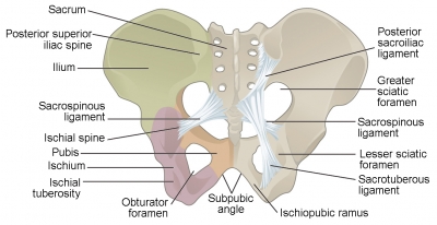
What is a haemorrhage?


Bleeding, also called hemorrhage, is the name used to describe blood loss. It can refer to blood loss inside the body, called internal bleeding, or to blood loss outside of the body, called external bleeding.


Blood loss can occur in almost any area of the body. Internal bleeding occurs when blood leaks out through a damaged blood vessel or organ. External bleeding happens when blood exits through a break in the skin


When blood from trauma irritates brain tissues, it causes swelling. This is known as cerebral edema. The pooled blood collects into a mass called a hematoma. These conditions increase pressure on nearby brain tissue, and that reduces vital blood flow and kills brain cells.


Bleeding can occur inside the brain, between the brain and the membranes that cover it, between the layers of the brain's covering or between the skull and the covering of the brain.


Picture Credit : Google


Which is the risk factor of stroke?


Lifestyle factors that increase your risk of stroke include high blood pressure, smoking, diabetes, high blood cholesterol levels, heavy drinking, high salt and high fat diet and lack of exercise. Someone who has already experienced a stroke is at increased risk of having another.


High blood pressure (hypertension) is the most significant risk factor for stroke. Blood pressure refers to the pressure inside the arteries. Normal blood pressure is around 120/80, while high-normal blood pressure is 120/80 to 140/90. High blood pressure is when your blood pressure is consistently over 140/90. This is called ‘hypertension’.


Hypertension means that the blood is exerting more pressure than is normal or healthy. Over time, this weakens and damages blood vessel walls, which can lead to stroke, particularly cerebral haemorrhage.


Hypertension may also cause thickening of the artery walls, resulting in narrowing and eventual blockage of the vessel (ischaemic stroke). In atherosclerosis, the pressure of your pumping blood could ‘hose off’ debris from damaged artery walls. The circulating debris (called emboli) can cause a stroke by lodging in and blocking a blood vessel in the brain.


Picture Credit : Google


Why does brain stroke happen?


A stroke, or "brain attack," occurs when blood circulation to the brain fails. Brain cells can die from decreased blood flow and the resulting lack of oxygen. There are two broad categories of stroke: those caused by a blockage of blood flow and those caused by bleeding into the brain. A blockage of a blood vessel in the brain or neck, called an ischemic stroke, is the most frequent cause of stroke and is responsible for about 80 percent of strokes. These blockages stem from three conditions: the formation of a clot within a blood vessel of the brain or neck, called thrombosis; the movement of a clot from another part of the body such as the heart to the brain, called embolism; or a severe narrowing of an artery in or leading to the brain, called stenosis. Bleeding into the brain or the spaces surrounding the brain causes the second type of stroke, called hemorrhagic stroke.


The most common symptoms of a stroke are:


  • Weakness or numbness of the face, arm, or leg on one side of the body

  • Loss of vision or dimming (like a curtain falling) in one or both eyes

  • Loss of speech, difficulty talking, or understanding what others are saying

  • Sudden, severe headache with no known cause

  • Loss of balance or unstable walking, usually combined with another symptom


Picture Credit : Google


What is the importance of water in our body?


The body needs water to stay alive. Every cell, tissue, and organ relies on a regular water supply to function properly. Water makes up more than half of the body. It is found inside cells, as well as in blood and other fluids, such as lymph, tears, saliva, sweat, and urine. The brain constantly monitors water level inside the body so it can make sure it maintains the correct balance.


Adequate water intake enables your body to excrete waste through perspiration, urination, and defecation. Water helps your kidneys remove waste from your blood and keep the blood vessels that run to your kidneys open and filter them out, according to the National Kidney Foundation. Water is also important for helping prevent constipation, points out the University of Rochester Medical Center. However, as research notes, there is no evidence to prove that increasing your fluid intake will cure constipation.


Your body loses fluids when you engage in vigorous exercise, sweat in high heat, or come down with a fever or contract an illness that causes vomiting or diarrhea, according to the Centers for Disease Control and Prevention. If you're losing fluids for any of these reasons, it's important to increase your fluid intake so that you can restore your body's natural hydration level. Your doctor may also recommend that you drink more fluids to help treat other health conditions, like bladder infections and urinary tract stones. If you're pregnant or nursing, you may want to consult with your physician about your fluid intake because your body will be using more fluids than usual, especially if you're breastfeeding.


Picture Credit : Google


What are glomeruli?


Inside the kidneys are thousands of tiny structures called glomeruli (one is called a glomerulus), working hard to filter toxic waste from the bloodstream. These tightly coiled clusters are the body’s smallest blood vessels (capillaries).


Each glomerulus works like a miniature sieve, transferring waste and excess water from the blood through the capillary wall into a renal tubule. This fluid drains away towards the bladder as urine.


The glomerulus receives its blood supply from an afferent arteriole of the renal arterial circulation. Unlike most capillary beds, the glomerular capillaries exit into efferent arterioles rather than venules. The resistance of the efferent arterioles causes sufficient hydrostatic pressure within the glomerulus to provide the force for ultrafiltration.


The glomerulus and its surrounding Bowman's capsule constitute a renal corpuscle, the basic filtration unit of the kidney.[2] The rate at which blood is filtered through all of the glomeruli, and thus the measure of the overall kidney function, is the glomerular filtration rate.


Picture Credit : Google


What is dialysis?


If the kidneys become damaged or diseased, a dialysis machine can be used to filter the blood instead. This large machine takes up to four times longer than kidneys to clean the body’s blood – which shows what an efficient unit the kidneys are.


The dialysis machine acts as an artificial set of kidneys. Blood flows from the body to the machine, toxic waste and excess fluid are removed, and cleaned blood is returned.


Some kinds of acute kidney failure get better after treatment. In some cases of acute kidney failure, dialysis may only be needed for a short time until the kidneys get better.


In chronic or end stage kidney failure, your kidneys do not get better and you will need dialysis for the rest of your life. If your doctor says you are a candidate, you may choose to be placed on a waiting list for a new kidney.


In hemodialysis, an artificial kidney (hemodialyzer) is used to remove waste and extra chemicals and fluid from your blood. To get your blood into the artificial kidney, the doctor needs to make an access (entrance) into your blood vessels. This is done by minor surgery to your arm or leg.


Picture Credit : Google


What is inside the kidney?


Each kidney has three layers – the outer layer (cortex), inner part (medulla), and central pelvis. Blood flows into the cortex for filtering. The medulla absorbs substances to return to the bloodstream. Waste is taken by tubes to the central pelvis, a collecting area where urine is emptied out into two tubes called ureters, and then passes to the bladder.


Multi-purpose organs


The two kidneys sit high in the back the abdomen. Each one is about the size of a fist, shaped like a bean, and surrounded by a protective layer of tissue.


Adrenal gland


The adrenal glands are small glands located on top of each kidney. They produce hormones that you can't live without, including sex hormones and cortisol. Cortisol helps you respond to stress and has many other important functions. Adrenaline released from this gland makes the heart beat faster in scary situations.


Renal artery


The renal arteries are responsible for carrying oxygen-rich blood to your kidneys, which in turn help the kidneys rid your body of waste and excess fluid. This artery carries blood into the kidney to be filtered.


Renal vein


The renal vein is an asymmetrically paired vessel that carries the deoxygenated blood from the kidney to the inferior vena cava. Cleaned blood is carried from the kidney by the renal vein.


Outer casing


The kidneys and adrenal glands are wrapped in a layer of fat and strong outer tissue. The outermost layer is a tough connective tissue layer called the renal fascia.


Central pelvis


Urine collects here, and is then sent to the bladder. The pelvis, which is shaped somewhat like a funnel that is curved to one side, is almost completely enclosed in the deep indentation on the concave side of the kidney, the sinus.


Renal cortex


The cortex is the outer part of the kidney. It contains the glomerulus and convoluted tubules.


The renal cortex is surrounded on its outer edges by the renal capsule, a layer of fatty tissue. Together, the renal cortex and capsule house and protect the inner structures of the kidney.


Renal medulla


This layer of the kidney absorbs water, making urine more concentrated. The renal medulla is split up into a number of sections, known as the renal pyramids. Blood enters into the kidney via the renal artery, which then splits up to form the interlobar arteries.


Left ureter


This is one of two tubes that carry urine down to the bladder. There are two ureters, one attached to each kidney. The upper half of the ureter is located in the abdomen and the lower half is located in the pelvic area. 


Nephrons


The kidneys contain tiny blood-filtering units called nephrons. Each nephron contains a glomerulus, a bundle of blood vessels surrounded by a capsule. As blood passes through the glomerulus, waste and excess water ooze into the capsule and are carried away by a tubule (tiny tube). Any useful substances, such as glucose, are absorbed by capillaries, while the waste is carried away to form urine.


Picture Credit : Google


What is the function of kidney?


Your two kidneys filter and clean the blood by removing toxic chemicals like the heart, the kidneys are at work every second of every day, producing a continuous flow of clean blood.


As blood circulates it picks up waste substances produced by the body's cells. These would poison you if they were not removed from the body. The kidneys extract the toxins and excess water from the blood and process them to make urine. As well as cleaning the blood, the kidneys also release hormones, stimulate red blood cell production and keep the body's water content balanced.


They release more urine if you have drunk a lot and less if you are dehydrated.


Picture Credit : Google


What is the difference between male and female pelvis?


The iliac crest of the male pelvis rises higher than the iliac crest of the female pelvis, and there is a greater distance between the anterior superior iliac spines of the female pelvis when compared to the male pelvis. This usually gives women a curvier appearance in the hip region, as compared to the average male. The female pelvic cavity is also going to be shallower, whereas the male pelvic cavity is deeper.


Although the female pelvis is wider than the typical male pelvis, these bones are thinner and lighter than the denser, rougher bones of the male pelvis.


The pelvic brim of the female is generally larger than the male’s inlet, which facilitates childbirth. The male inlet is smaller and heart-shaped, and a trick to remembering that is to remember the following phrase: the way to a man’s heart is through his pelvis!


The sacrum of the female is shorter, wider, and has a greater curve, whereas the male sacrum is thinner, longer, and is less curved. The coccyx bone of the male curves more toward the front of the body in comparison to the female’s coccyx.


Picture Credit : Google


What is the anatomy of pelvis?


Pelvic girdle


The pelvis is made up of two hip bones, one on each side. Each hip bone has three parts - the ilium, ischium, and pubis. They connect to the sacrum - the lower  part of the spine - to form a ring like shape, called the pelvic girdle.


Spine


The spine, or backbone is a column of bones that runs from the pelvis to the neck. The spine is classified into 4 distinct areas.


The cervical area consists of 7 bony parts in the neck. The thoracic spine consists of 12 bony parts in the back area. The lumbar spine consists of 5 bony segments in the lower back area; 5 sacral* bones; and 4 coccygeal* bones (the number of coccygeal bones can vary from 3 to 5). (* By adulthood, the 5 sacral vertebrae fuse to form 1 bone, and the 4 coccygeal vertebrae fuse to form 1 bone.)


Sacrum


At the base of the spine, this triangular-shaped bone connects the two hip bones. It consists of the last four or five vertebrae that by adulthood, fuse together to form a single bone. Located just above the coccyx and wedged between the right and left iliac bones (hip bones), the sacrum forms the back wall of the pelvis. The coccyx, commonly referred to as the tailbone, is the very bottom segment of the vertebral column. The right and left iliac bones are joined together in the front by the symphysis pubis.


Ilium


The largest bone in the pelvis is the ilium, or hip bone. One on each side connects the muscles used to stand and walk. The body of the ilium forms the superior part of the acetabulum (acetabular roof). Immediately above the acetabulum, the ilium expands to form the wing (or ala).


Fixed joint


These bones are held firmly together by strong ligaments. The relatively fixed joint between the sacrum and the lumbar spine is ideal for bearing the load of the upper body. At the most ventral area of the pelvis, there is a fibrocartilaginous joint in the pubic symphysis.


Pelvic cavity


The pelvic cavity is formed by three bilateral pairs of bones (pubis, ilium and ischium) and two posteriorly located bones (sacrum and coccyx). The intestines and bladder are contained here, surrounded by the protective pelvis.


Pubis


The pubis is one of the two smallest bones in the pelvis. In vertebrates, the pubic bone is the most forward-facing (ventral and anterior) of the three main bones making up the pelvis. The left and right pubic bones are each made up of three sections, a superior ramus, inferior ramus, and a body.


Pubic symphysis


This strong cartilage joint connects the two pubis bones together. It is located in front of and below the urinary bladder. In males, the suspensory ligament of the penis attaches to the pubic symphysis.


Coccyx


Below the sacrum is the Coccyx, or tailbone all that remains of the tail of our distant ancestors. Although the tailbone is considered vestigial (or no longer necessary) in the human body, it does have some function in the pelvis. For instance, the coccyx is one part of a three-part support for a person in the seated position. Weight is distributed between the bottom portions of the two hip bones (or ischium) and the tailbone, providing balance and stability when a person is seated.


Hip joint


The ball shaped top of the thighbone sits in this hollow creating the ball and socket hip joint. The adult os coxae, or hip bone, is formed by the fusion of the ilium, the ischium, and the pubis, which occurs by the end of the teenage years. The 2 hip bones form the bony pelvis, along with the sacrum and the coccyx, and are united anteriorly by the pubic symphysis.


Ischium


The lowest bone in the pelvis the ischium carries all the weight when the body is sitting down. Situated below the ilium and behind the pubis, it is one of these three bones whose fusion creates the hip. The superior portion of this bone forms approximately one third of the acetabulum.


Holes


Small holes in the bones are for nerves and blood vessels.


Picture Credit : Google