How does the stomach work?

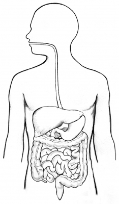
The stomach is a muscular hollow organ. It takes in food from the esophagus (gullet or food pipe), mixes it, breaks it down, and then passes it on to the small intestine in small portions.

The entire digestive system is made up of one muscular tube extending from the mouth to the anus. The stomach is an enlarged pouch-like section of this digestive tube. It is located on the left side of the upper abdomen and shaped somewhat like an oversized comma, with its bulge pointing out to the left. The stomach’s shape and size vary from person to person, depending on things like people’s sex and build, but also on how much they eat.

At the point where the esophagus leads into the stomach, the digestive tube is usually kept shut by muscles of the esophagus and diaphragm. When you swallow, these muscles relax and the lower end of the esophagus opens, allowing food to enter the stomach. If this mechanism does not work properly, acidic gastric juice might get into the esophagus, leading to heartburn or an inflammation.

The upper-left part of the stomach near the opening curves upward towards the diaphragm. This part is called fundus. It is usually filled with air that enters the stomach when you swallow. In the largest part of the stomach, called the body, food is churned and broken into smaller pieces, mixed with acidic gastric juice and enzymes, and pre-digested. At the exit of the stomach, the body of the stomach narrows to form the pyloric canal, where the partially digested food is passed on to the small intestine in portions.

The stomach wall is made up of several layers of mucous membrane, connective tissue with blood vessels and nerves, and muscle fibers. The muscle layer alone has three different sub-layers. The muscles move the contents of the stomach around so vigorously that solid parts of the food are crushed and ground, and mixed into a smooth food pulp.

The inner mucous membrane (lining) has large folds that are visible to the naked eye. These folds run toward the exit of the stomach, providing “pathways” along which liquids can quickly flow through the stomach. If you look at the mucous membrane under a microscope, you can see lots of tiny glands. There are three different types of glands. These glands make digestive enzymes, hydrochloric acid, mucus and bicarbonate.

Gastric juice is made up of digestive enzymes, hydrochloric acid and other substances that are important for absorbing nutrients – about 3 to 4 liters of gastric juice are produced per day. The hydrochloric acid in the gastric juice breaks down the food and the digestive enzymes split up the proteins. The acidic gastric juice also kills bacteria. The mucus covers the stomach wall with a protective coating. Together with the bicarbonate, this ensures that the stomach wall itself is not damaged by the hydrochloric acid.

Credit : NCBI 

Picture Credit : Google 

Which is the smallest muscle in human body?

Did you know the smallest muscle in the body is located in the ear? Called the stapedius, it is said to be less than 2 mm long. It supports the smallest bone in the body, called the stapes, which is part of the middle ear and helps conduct vibrations to the inner ear. Its purpose is to stabilise the smallest bone in the body.

The stapedius dampens the vibrations of the stapes by pulling on the neck of that bone. As one of the muscles involved in the acoustic reflex it prevents excess movement of the stapes, helping to control the amplitude of sound waves from the general external environment to the inner ear.

If there is damage to the nerve to stapedius, wider oscillations of the stapes will occur resulting in hyperacusis—sounds being perceived as extremely loud, more so than they actually are to a person without damage to this nerve.

Picture Credit : Google 

What is a stroke?

 Stroke is a medical condition in which blood supply is severely reduced to parts of the brain resulting in cell death. A stroke occurs when a blood vessel in the brain ruptures and bleeds, or when there’s a blockage in the blood supply to the brain. The rupture or blockage prevents blood and oxygen from reaching the brain’s tissues. Without oxygen, brain cells and tissue become damaged and begin to die within minutes. According to the Centers for Disease Control and Prevention (CDC)Trusted Source, stroke is a leading cause of death in the United States. Every year, more than 795,000 U.S. people have a stroke. There are three primary types of strokes:

Transient ischemic attack (TIA) involves a blood clot that typically reverses on its own.
Ischemic stroke involves a blockage caused by either a clot or plaque in the artery. The symptoms and complications of ischemic stroke can last longer than those of a TIA, or may become permanent.
Hemorrhagic stroke is caused by either a burst or leaking blood vessel that seeps into the brain.

Stroke symptoms can include: ,paralysis numbness or weakness in the arm, face, and leg, especially on one side of the body, trouble speaking or understanding others, slurred speech,confusion, disorientation, or lack of responsiveness, sudden behavioral changes, especially increased agitation, vision problems, such as trouble seeing in one or both eyes with vision blackened or blurred, or double vision, trouble walking, loss of balance or coordination, dizziness, severe, sudden headache with an unknown cause,  seizures, nausea or vomiting

Proper medical evaluation and prompt treatment are vital to recovering from a stroke. According to the American Heart Association and American Stroke Association, “Time lost is brain lost.”

Credit : Healthline

Picture Credit : Google

Brain triggers, cues and planned movement

When we are confronted with a red signal, we wait till it turns green before we make any movement - be it crossing the road on foot or riding a vehicle. In any sprinting race, the starters-both elite athletes in the international stage and youngsters in school-level races - wait for a signal before bursting from the starting line to race to the finish.

Our brain has planned our precise movements but waits for the execution until a specific cue. Scientists from the Max Planck Florida Institute for Neuroscience, HHMI's Janelia Research Campus, the Allen Institute for Brain Science, and others have discovered the brain network that responds to a cue by turning plans into action.

Brain is like an orchestra

One of the authors of the paper, published in the scientific journal Cell in March, suggests that the brain is like an orchestra. Just like how an orchestra needs a conductor to ensure a perfect symphony, the brain too has areas that act as a conductor and ensure that plans are converted into action at exactly the right time. By simultaneously recording the activity of hundreds of neurons when a mouse performed a cue-triggered movement task, the team was able to identify the neural circuits that serve as the conductor. The task involved the mice licking to the right if whiskers were touched or to the left if whiskers were not touched. The mice not only had to get it correct, but also had to delay their movement until a go cue was played in order to receive a reward.

The scientists were able to correlate complex neuronal activity to various stages of the tasks. They next identified a circuit of neurons in which brain activity occurred after the go cue, leading to the execution. By using optogenetics to activate or inactivate this circuit of neurons in the brain while performing additional tasks, the researchers were able to confirm their discovery.

Can it improve mobility?

 Apart from serving as fundamental advances in our understanding of how our brain functions, this discovery could also have important clinical implications. People who have had an accident or experiencing motor disorders sometimes have difficulty in self-initiated movement. Environmental cues, both visual and auditory, could well trigger movements that can improve the person's mobility dramatically. 

This phenomenon wherein different mechanisms are employed for self-initiated and cue-triggered movements is known as paradoxical kinesia. Understanding how our brain functions during cue-triggered movements may help us in treatments.

Picture Credit : Google 

Cluster Headache Awareness Day (March 21)

The 21st of March is the “Cluster Headache Awareness Day”, a prominent event to promote CH on scientific and public levels. The spring equinox represents the perfect choice for a disease with such a great circadian and circannual  rhythmicity. Indeed, a vast majority of CH subjects experienced a CH reactivation during the seasonal shift in spring and autumn; in some cases, the circannual timing becomes so scheduled that patients do not plan activities and slowly slide toward social withdrawal just for the fear of a novel cluster period. The 21st of March is well remembered by CH patients, because starting from this date when the daylight increases there are positive effects for patients with night attacks.

With a prevalence of 0.12%, Cluster Headache (CH) is the most frequent trigeminal autonomic cephalalgia. CH is characterised by a typical clinical picture, namely a strictly unilateral, very severe, headache lasting 15 to 180 minutes associated with prominent cranial autonomic features, which are lateralized and ipsilateral to the headache.

In its episodic subtype (85% of CH subjects), CH attacks are present only in limited period of the years, lasting weeks to months (the so-called cluster periods), alternating with remission phases of at least 3 months of duration. By contrast, in the chronic CH subtype the remissions are shorter or not present at all, and the burden on the individual becomes not imaginable.

During the day, CH subjects may experience several attacks during day, often distributed during the night and with a typical circadian rhythm.

People suffering from CH consistently report severe limitations in activities of daily living and social-activity participation. Nonetheless, their working activity and career may be hindered.

CH has historically been considered as “rare”. If it is true that its prevalence falls far below migraine, it is also true that CH does not represent a rare disease. All in all, the direct and indirect (work absenteeism, sick leave, and so on) costs make CH a burden not only for the individual but for the society globally.

Therapeutic options are still limited, with most of the preventive medications being non-CH specific and borrowed from other medical conditions. Subcutaneous sumatriptan and high-flow oxygen represent the first-line choices for the acute management of CH pain. A novel anti-CGRP monoclonal antibody, which proved effective in migraine, has been approved for the preventive treatment of episodic CH, but not for the most severe chronic CH subtype. Long-term observation is needed to confirm the real-life impact of these novel drugs, hoping for a novel and specific alternative to treat CH.

Finally, CH is still little known outside the headache centres leading to diagnostic delay, low quality of counselling to the patients and sub-optimal therapeutic management.

Credit : International Headache Society 

Picture Credit : Google