What is spinal cord?


The spinal cord is the body’s information superhighway, along which billions of nerve cells carry messages that enable the body to move and function.


It has two main sections: a butterfly-shaped inner mass of grey matter surrounded by outer white matter. White matter is made up of nerve fibre that relay signals to and from the brain. Grey matter contains neurons that receive signals from receptors around the body, and send on instructions to the muscles.


When the body moves, messages travel from the brain down the spinal cord. Messages also are carried up the spinal cord to the brain so a person can feel sensations.


There are nerves that branch off the spinal cord. They are called spinal nerves. The nerves are divided into five main sections (from top to bottom): cervical, thoracic, lumbar, sacral, and coccygeal. The spinal cord and nerves are very fragile.


Picture Credit : Google


What are the regions of the backbone?


The vertebrae that make up the backbone are often divided into five regions:


Cervical vertebrae


The seven cervical vertebrae support the head, with the top two vertebrae, the atlas and axis, enabling the head to nod and turn.


Thoracic vertebrae


The 12 thoracic vertebrae connect with the ribs, forming the back of the ribcage. Each thoracic vertebra has two ‘demi facets,’ superiorly and inferiorly placed on either side of its vertebral body. The demi facets articulate with the heads of two different ribs.


Lumbar vertebrae


There are five lumbar vertebrae, which support most of the body’s weight. Lumbar vertebrae have very large vertebral bodies, which are kidney-shaped. They lack the characteristic features of other vertebrae, with no transverse foramina, costal facets, or bifid spinous processes.


Fused bones of the sacrum


A group of five bones connect the backbone to the pelvic girdle. It is described as an inverted triangle, with the apex pointing inferiorly. On the lateral walls of the sacrum are facets for articulation with the pelvis at the sacro-iliac joints.


Fused bones of the coccyx


These four bones are an attachment point for muscles, tendons, and ligaments. It is recognised by its lack of vertebral arches. Due to the lack of vertebral arches, there is no vertebral canal.


Picture Credit : Google


What are spinal joints?


The spurs of bone on the back of each vertebra slot together to form joints that glide back and forth as the spine moves, called facet joints. The shapes of the bones limit how much each joint can move. Discs of cartilage between the bones absorb shock by squashing slightly. They also stop the bones from grinding painfully against each other as they move.


Facet joints appear to have little influence on the range of side bending (lateral flexion). These functions can be disrupted by degeneration, dislocation, fracture, injury, instability from trauma, osteoarthritis, and surgery. In the thoracic spine the facet joints function to restrain the amount of flexion and anterior translation of the corresponding vertebral segment and function to facilitate rotation. Cavitation of the synovial fluid within the facet joints is responsible for the popping sound (crepitus) associated with manual spinal manipulation, commonly referred to as "cracking the back."


The facet joints, both superior and inferior, are aligned in a way to allow flexion and extension, and to limit rotation. This is especially true in the lumbar spine.


Picture Credit : Google


What is the anatomy of backbone?


To ensure the spinal column is well protected, the joints between each vertebra only allow limited movement. But all the small movements adds up, allowing the spine to bend backwards and forwards and side to side, as well as twist and turn.


Hip bone


The left and right hip bones (innominate bones, pelvic bones) are two irregularly shaped bones that form part of the pelvic girdle – the bony structure that attaches the axial skeleton to the lower limbs.


Coccyx (tailbone)


The coccyx is a triangular arrangement of bone that makes up the very bottom portion of the spine below the sacrum. It represents a vestigial tail, hence the common term tailbone. These fused bones help to bear the body’s weight when sitting.


Shock absorbing disc


An intervertebral disk acts as shock absorber between each of the vertebrae in the spinal column by keeping the vertebrae separated when there is impact from activity. They also serve to protect the nerves that run down the middle of the spine and intervertebral disks.These plates of cartilage are sandwiched between the vertebrae.


Vital protection


The area between the spinal cord and the vertebrae is called the epidural space. This contains blood vessels and connective tissue that forms a protective cushion around the spinal cord. Nerves gain access to the spinal cord through gaps in the bones, called intervertebral foramina.


Picture Credit : Google


What is the function of backbone?


The backbone, also called the spine, runs down the back of the body, from the base of the skull to the coccyx. It provides strong support for the head and body, while allowing the body to twist and bend. It also protects the spinal cord – the thick bundle of nerves that carries messages between the brain and the body.


The human backbone is formed by small, pillar – shaped bones called vertebrae (one is called a vertebra). These stacks together to form a strong, flexible, S-shaped column. This shape makes the backbone springy enough to absorb shock during movement, while larger vertebrae in the lower back help to support the upper body’s weight. Each vertebra slots into its neighbour to form a flexible but secure tunnel surrounding the spinal cord.


Picture Credit : Google


Which muscles are important for maintaining posture?


The layer of stabilizing back muscles plays an important role in a baby’s developing ability to sit and move around independently.


Push-ups


Babies begin to build strength in their stabilizing muscles from about three months old. They lie on their tummies and lift their arms in the air to flex these muscles.


Sitting up                 


By nine months old, most babies have developed their stabilizing muscles enough to be able to sit up on their own.


First steps


At around a year old, a baby can stand on her feet and start to walk without help. Stabilizing muscles are still gaining strength, so early attempts can be wobbly.


Picture Credit : Google


What is the anatomy of core muscles?


The core muscles support the spine and provide stability.


Serratus posterior superior


The serratus posterior muscles extend obliquely from the vertebral column to the rib cage. During breathing, this muscle helps lift the ribs.


Rhomboid major


The rhomboids are two bilateral, superficial muscles located in the upper back. This muscle links the shoulder blade to the ribcage.


Erector spinae


The erector spinae muscles comprise the intermediate layer of the deep (intrinsic) muscles of the back This group of three muscles forms a supporting column on either side of the spine.


Serratus posterior inferior


The serratus posterior inferior muscle is one of the back’s two intermediate muscles (the other is called the serratus posterior superior muscle), which means it’s neither a surface nor a deep muscle. During breathing, this muscle helps pull the lower ribs down.


External intercostals muscle


The external intercostal muscles are the most superficial set of muscles that occupy the 11 intercostal spaces.  This pulls the ribcage up and out when breathing in. This is one of a set of muscles that raises the ribs and expands the chest.


Rotators


These small muscles run all the way up either side of the spine. Its function is related to the glenohumeral joint, where the muscles of the cuff function both as the executors of the movements of the joint and the stabilization of the joint as well.


Psoas major


Psoas major is a long, thick, fusiform muscle located in the lumbar region of the trunk lateral to the lumbar vertebrae and medial to the quadratus lumborum muscle. This muscle helps to move the hip.


Gluteal muscles


The gluteus maximus is the most superficial gluteal muscle that forms the prominence of the gluteal region. These muscles stabilize the hip, pelvis, and back.


Multifidus


Multifidus is a group of short, triangular muscles that along with the semispinalis and rotatores comprises the transversospinal group of deep back muscles. The joints of the lower back are stabilized by this long, thin muscle.


Picture Credit : Google


What muscles help support the spine?


The muscles of the neck and back provide a strong support system for the spine – the long line of interlocking bones that helps to keep the upper body stable and upright. Some muscles also help with breathing by lifting and lowering the ribs.


Back muscles pull the spine backwards, bend it sideways, or rotate it, allowing the back to perform a wide range of bending and turning movements. Layers of muscle packed around the spine also protect it against injury from pressure or knocks.


The back has three main muscle layers, which work together to stabilize and move the torso, and help with breathing. The deeper layer sometimes called the core muscles, hold the body upright and stop you flopping forward when you bend at the waist.


Picture Credit : Google


What is the anatomy of chest muscles?


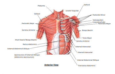
The muscles of the chest help with the process of breathing. They pull the ribs up and out, making more space for the lungs to expand as we breathe in. When they relax, the space gets smaller and the air is forced out again.


Clavicle


The clavicle is an elongated, S-shaped bone that rests horizontally at the sternum across the upper part of the ribcage, and the acromial end of the scapula. This is also called collarbone.


Pectoralis major


The largest chest muscle, it is attached to the sternum, clavicle, humerus (upper arm bone), and ribs. The pectoralis major has a broad origin, based on which it is divided into three parts: clavicular part, sternocostal part and abdominal part. All three parts converge laterally and insert onto the greater tubercle of humerus.


Sternum


The sternum is the bone that lies in the anterior midline of our thorax. It forms part of the rib cage and the anterior-most part of the thorax. Several muscles attach to the sternum, or breastbone.


Deltoid


The deltoid is a thick, triangular shoulder muscle. It gets its name because of its similar shape to the Greek letter ‘delta’ (?). Covering the shoulder joint, this muscle raises the arm.


Serratus anterior


The serratus anterior muscle is a fan-shaped muscle at the lateral wall of the thorax. Its main part lies deep under the scapula and the pectoral muscles. This muscle connects to the upper ribs.


Costal cartilage


Tough, springy tissue connects the sternum to the ribs. The costal cartilage is segments of cartilage that connect the sternum to the ribs and help to extend the ribs into a forward motion.  This cartilage also contributes to elasticity within the walls of the thorax, allowing the chest to expand during respiration


Rectus abdominis


Rectus abdominis, informally known as the abs muscle, is a long muscle of the anterior abdominal wall. Attached to the lower sternum and coastal cartilages, this helps keep the body upright.


External oblique


External abdominal oblique is a paired muscle located on the lateral sides of the abdominal wall. Along with internal abdominal oblique and transversus abdominis, it comprises the lateral abdominal muscles. This outer muscle layer helps to force air out of the lungs when we breathe out.


Rib


Each rib is thin and curved, with an inside groove for veins, arteries, and nerves. In most tetrapods, ribs surround the chest, enabling the lungs to expand and thus facilitate breathing by expanding the chest cavity.


Internal oblique muscles


Internal abdominal oblique is a broad thin muscular sheet found on the lateral side of the abdomen. These help to push air out of the lungs when we breathe out.


Intercostals muscles


Between the ribs are three layers of intercostals muscles (“intercostals” simply means “between the ribs”). The muscle fibres run in different directions so the ribs can be pulled in different ways.


Internal muscle


Internal intercostals are part of the muscles of the thoracic wall located in the intercostal spaces between the ribs. When you breathe out, this muscle pulls the ribcage down and out.


External muscle


The external intercostal muscles are the most superficial set of muscles that occupy the 11 intercostal spaces.  This pulls the ribcage up and out when breathing in.


Innermost muscle


Innermost intercostals comprise the third and deepest layer of intercostal muscles. This muscle lowers the ribs when breathing out.


Picture Credit : Google


What is the function of ribs and chest muscles?


The chest, or thorax, lies between the neck and the abdomen. Inside the thorax lie the heart, lungs, and major blood vessels. The ribcage surrounding them is formed by the backbone, ribs, costal cartilages, and sternum (breastbone).


The ribcage is strong enough to protect the vital organs, but flexible enough to expand and contract for breathing. Attached to the ribcage are the muscles of the chest. Together with the diaphragm, many of these muscles help with breathing.


They move superiorly, inferiorly, anteriorly and posteriorly to facilitate breathing (their flexibility in their movement increases/decreases the size of the thoracic cavity; assisting the lungs in respiration. Control of these movements is via the diaphragm, external intercostals, and the intercartilaginous portion of the internal intercostals). They play a role in erythropoiesis during development (at birth, the erythropoiesis sites change, it recedes in long bones and persists in flat bones, like ribs).


Picture Credit : Google


What is the function of chest and back?


The chest houses the two powerhouse organs that keep the body running – the heart and lungs. All the body’s cells are supplied with essential blood and oxygen thanks to these vital organs. The backbone supports the body and protects the spinal column, which carries messages to and from the brain.


The chest wall is comprised of skin, fat, muscles, and the thoracic skeleton. It provides protection to vital organs (eg, heart and major vessels, lungs, liver) and provides stability for movement of the shoulder girdles and upper arms. 


One important organ in the chest is the thymus, a small butterfly-shaped organ located between the heart and the sternum, or breastbone. This organ belongs to the immune system, and its job is to produce T cells, a type of white blood cell. These are formally known as T lymphocytes; the “T” stands for thymus, where the cells originate.


Picture Credit : Google


What part of your body helps you keep your balance?


Different body systems work to keep you balanced. Signals from the inner ear combine with visual signals from the eyes, pressure sensors in the skin, and stretch sensors in the muscles to reveal the body’s position. The brain processes this and makes any adjustments to stop the body falling over.


Utricle and saccule


Inside the inner ear are two tiny organs that sense movements of the head in a straight line. The utricle detects forward and backward movement, and the saccule detects up-and-down movement.


Forward and backward movement, such as travelling in a car, is detected by the utricle. Up-and-down motion, such as riding in a lift, is sensed by the saccule.


Picture Cedit : Google


What are the inner parts of the ear?


Inner ear shows the semicircular canals and the cochlea. The three fluid-filled canals sit at right angles to each other. When you move your head, the fluid swishes around and causes tiny hairs to bend. This sends nerve signals to the brain, which works out the direction in which you are moving. The snail-shaped cochlea converts sounds into nerve impulses.


Inner ear


The semicircular canals and cochlea are part of the inner ear. They sit within a hollow in the temporal bone of the skull.


Inside a semicircular canal


Bulb-shaped areas called ampullae sit at the base of each semicircular canal. In the middle of each ampulla is the cupula, which houses the bundle of movement-sensing hairs. The brain co-ordinates feedback from the ampullae to maintain a constant fix on the body’s position so the body can keep it balanced.


Utricle


The utricle is a small membranous sac (part of the membranous labyrinth) and paired with the saccule lies within the vestibule of the inner ear. It has an important role in orientation and static balance, particularly in horizontal tilt. The utricle is sensitive to forward and backward movements.


Saccule


The saccule is a small membranous sac, paired with the utricle, within the vestibule of the inner ear. It is part of the membranous labyrinth and has an important role in orientation and balance, particularly in vertical tilt. The saccule senses vertical movements of the head.


Oval window


The stapes, or stirrup bone, fits here to pass sound waves to the cochlea. Sound waves cause vibration of the tympanic membrane and the ossicles transmit those vibrations to the oval window, which leads to movement of fluid within the cochlea and activation of receptors for hearing.


Cochlea


The cochlea contains the spiral organ of Corti, which is the receptor organ for hearing. It consists of tiny hair cells that translate the fluid vibration of sounds from its surrounding ducts into electrical impulses that are carried to the brain by sensory nerves. This snail-shaped organ turns vibrations into audible sounds.


Auditory nerve


This nerve carries signals from the ear to the brain.  It is one of the many pieces that make up the auditory system, which enables effective hearing.


Organ of Corti


Running through the middle of the cochlea is the organ of Corti, the main receptor for hearing. Sound waves create vibrations that make wave-like movements in the fluid inside the organ of Corti. These bend the hairs, producing nerve signals, which are sent to the brain to be registered as sounds.


Picture Credit : Google


How do ears help with balancing and hearing?


As well as providing our sense of hearing, the ears help us to keep our balance and send vital information to the brain when we move.


The inner ear is the part deepest inside the head. It contains three fluid-filled tubes called semicircular canals. As we move, the fluid inside the canals moves, sending messages to the brain to help us keep our balance. Also in the inner ear is the cochlea, which converts sounds to hearing.


Information coming from the vestibular system is processed in the brain and then sent on to other organs that need this information, such as the eyes, joints or muscles. This allows us to keep our balance and know what position our body is in.


In some situations, for example on a ship or airplane, different sensory organs (e.g. the eyes and the organ of balance) send contradictory messages to the brain. This can cause us to feel unwell, dizzy or nauseous.


The vestibular system is especially sensitive in children, and reacts more slowly to movements as we grow older. Inner ear infections and other problems may also affect how well our sense of balance works.


Picture Credit : Google


What is the ear in anatomy?


Pinna (outer ear)


The pinna is the only visible part of the ear (the auricle) with its special helical shape. It is the first part of the ear that reacts with sound. The function of the pinna is to act as a kind of funnel which assists in directing the sound further into the ear. This flap channels sounds into the ear canal.


Ear canal


The ear canal, also called the external acoustic meatus, is a passage comprised of bone and skin leading to the eardrum. The ear is comprised of the ear canal (also known as the outer ear), the middle ear, and the inner ear.


Eardrum


The eardrum is a thin, film, about 9 mm (0.4 in) wide, which sits at the entrance to the middle ear and vibrates when sound waves hit it. The eardrum also helps to stop debris from getting inside the ear and damaging it.


Hammer (malleus)


Vibrations from the eardrum are picked up by this bone. The malleus is a bone situated in the middle ear. It is the first of the three ossicles, and attached to the tympanic membrane. The head of the malleus is the large protruding section, which attaches to the incus. The head connects to the neck of malleus, and the bone continues as the handle of malleus, which connects to the tympanic membrane. Between the neck and handle of the malleus, lateral and anterior processes emerge from the bone.[2


Stirrup (stapes)


The tiny stirrup bone vibrates and moves the oval window in the cochlea. If the stapes becomes damaged, such as from severe head trauma, a person may lose some or all of their ability to hear. Because the ossicles are a chain of bones, this also holds true for the incus and malleus.


Semicircular canals


These three fluid-filled tubes contain sensors that detect movement. Your semicircular canals are three tiny, fluid-filled tubes in your inner ear that help you keep your balance. When your head moves around, the liquid inside the semicircular canals sloshes around and moves the tiny hairs that line each canal


Cochlea


The snail-shaped cochlea is filled with liquid and lined with tiny hair cells that detect vibrations. The cochlea interacts with the middle ear via two holes that are closed by membranes: the oval window, which is located at the base of the scala vestibuli and which undergoes pressure from the stapes (see ‘middle ear’), and the round window, which seals the base of the tympanic membrane and is used to relieve pressure.


Hair cells


Each hair cell in the cochlea is topped by groups of microscopic hairs. Incoming vibrations bend the hairs by different amounts. These vibration patterns are turned into nerve messages and sent to the brain.


Anvil (incus)


The incus lays at the center of the ossicles, connecting the malleus to the stapes. It is shaped like an anvil, which is why ‘the anvil’ is a widely used alternative name for the bone. Vibrations from the hammer to the stirrup are transmitted through the anvil.


Smallest bones


The ear contains three of the tiniest bones in the human body. The stirrup bone is the smallest of all, at about the size of a grain of rice.


Picture Credit : Google